 Movie
Movie Controller


+ Open data

- Basic information
| Entry | 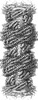 | ||||||||||||
|---|---|---|---|---|---|---|---|---|---|---|---|---|---|
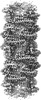
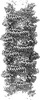
| Title | DpHF7 filament | ||||||||||||
 Map data Map data | DpHF7 filament | ||||||||||||
 Sample Sample |
| ||||||||||||
 Keywords Keywords |  Filament / pH / designed / DE NOVO PROTEIN Filament / pH / designed / DE NOVO PROTEIN | ||||||||||||
| Biological species | synthetic construct (others) | ||||||||||||
| Method | helical reconstruction / cryo EM / Resolution: 3.3 Å | ||||||||||||
 Authors Authors | Lynch EM / Farrell D / Shen H / Kollman JM / DiMaio F / Baker D | ||||||||||||
| Funding support |  United States, 3 items United States, 3 items
| ||||||||||||
 Citation Citation | Journal: Nat Nanotechnol / Year: 2024 Title: De novo design of pH-responsive self-assembling helical protein filaments. Authors: Hao Shen / Eric M Lynch / Susrut Akkineni / Joseph L Watson / Justin Decarreau / Neville P Bethel / Issa Benna / William Sheffler / Daniel Farrell / Frank DiMaio / Emmanuel Derivery / James ...Authors: Hao Shen / Eric M Lynch / Susrut Akkineni / Joseph L Watson / Justin Decarreau / Neville P Bethel / Issa Benna / William Sheffler / Daniel Farrell / Frank DiMaio / Emmanuel Derivery / James J De Yoreo / Justin Kollman / David Baker /  Abstract: Biological evolution has led to precise and dynamic nanostructures that reconfigure in response to pH and other environmental conditions. However, designing micrometre-scale protein nanostructures ...Biological evolution has led to precise and dynamic nanostructures that reconfigure in response to pH and other environmental conditions. However, designing micrometre-scale protein nanostructures that are environmentally responsive remains a challenge. Here we describe the de novo design of pH-responsive protein filaments built from subunits containing six or nine buried histidine residues that assemble into micrometre-scale, well-ordered fibres at neutral pH. The cryogenic electron microscopy structure of an optimized design is nearly identical to the computational design model for both the subunit internal geometry and the subunit packing into the fibre. Electron, fluorescent and atomic force microscopy characterization reveal a sharp and reversible transition from assembled to disassembled fibres over 0.3 pH units, and rapid fibre disassembly in less than 1 s following a drop in pH. The midpoint of the transition can be tuned by modulating buried histidine-containing hydrogen bond networks. Computational protein design thus provides a route to creating unbound nanomaterials that rapidly respond to small pH changes. | ||||||||||||
| History |
|
- Structure visualization
| Supplemental images |
|---|

- Downloads & links
-EMDB archive
| Map data | emd_42075.map.gz | 32.6 MB |  EMDB map data format EMDB map data format | |
|---|---|---|---|---|
| Header (meta data) | emd-42075-v30.xmlemd-42075.xml | 15 KB 15 KB | Display Display | EMDB header |
| FSC (resolution estimation) | emd_42075_fsc.xml | 12.7 KB | Display | FSC data file |
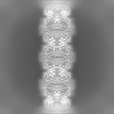
| Images |  emd_42075.png emd_42075.png | 96.4 KB | ||
| Filedesc metadata | emd-42075.cif.gz | 5.6 KB | ||
| Others | emd_42075_half_map_1.map.gzemd_42075_half_map_2.map.gz | 200.1 MB 200.1 MB | ||
| Archive directory |  http://ftp.pdbj.org/pub/emdb/structures/EMD-42075ftp://ftp.pdbj.org/pub/emdb/structures/EMD-42075 http://ftp.pdbj.org/pub/emdb/structures/EMD-42075ftp://ftp.pdbj.org/pub/emdb/structures/EMD-42075 | HTTPS FTP |
-Related structure data
| Related structure data | 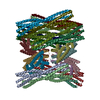 8ub3MC 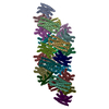 8uaoC 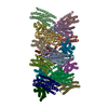 8ubgC M: atomic model generated by this map C: citing same article ( |
|---|
-Links
| EMDB pages | EMDB (EBI/PDBe) / EMDataResource |
|---|
-Map
| File | Download / File: emd_42075.map.gz / Format: CCP4 / Size: 216 MB / Type: IMAGE STORED AS FLOATING POINT NUMBER (4 BYTES) | ||||||||||||||||||||
|---|---|---|---|---|---|---|---|---|---|---|---|---|---|---|---|---|---|---|---|---|---|
| Annotation | DpHF7 filament | ||||||||||||||||||||
| Voxel size | X=Y=Z: 1.05 Å | ||||||||||||||||||||
| Density |
| ||||||||||||||||||||
| Symmetry | Space group: 1 | ||||||||||||||||||||
| Details | EMDB XML:
|
-Supplemental data
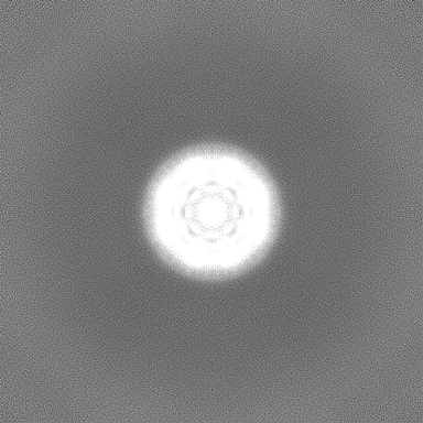
-Half map: Half Map 1
| File | emd_42075_half_map_1.map | ||||||||||||
|---|---|---|---|---|---|---|---|---|---|---|---|---|---|
| Annotation | Half Map 1 | ||||||||||||
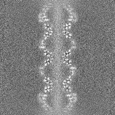
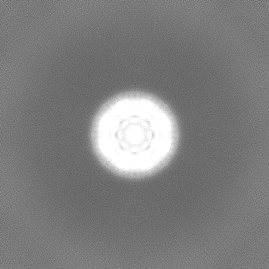
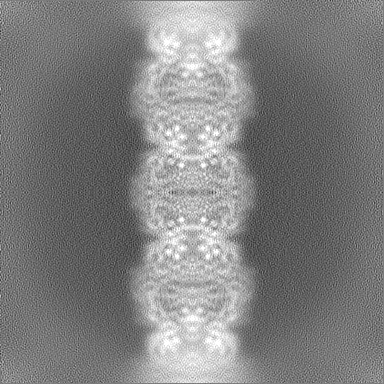
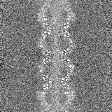
| Projections & Slices |
| ||||||||||||
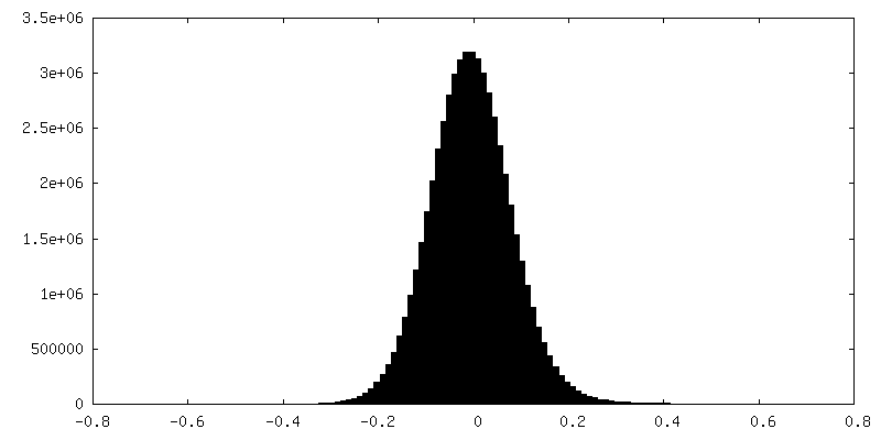
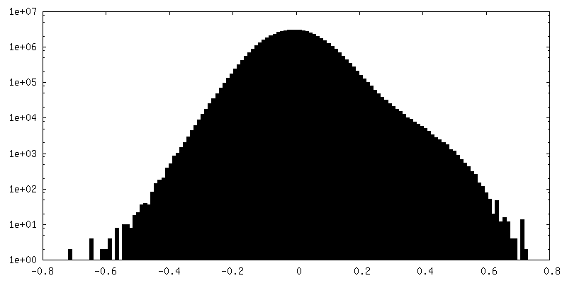
| Density Histograms |
 Z
Z Y
Y X
X
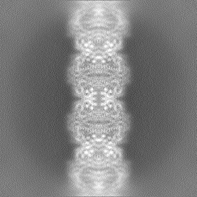

-Half map: Half Map 2
| File | emd_42075_half_map_2.map | ||||||||||||
|---|---|---|---|---|---|---|---|---|---|---|---|---|---|
| Annotation | Half Map 2 | ||||||||||||
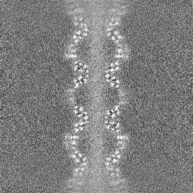
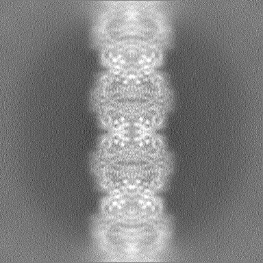
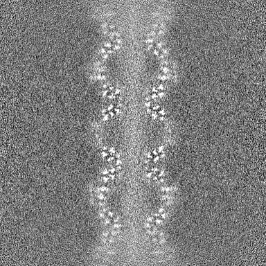
| Projections & Slices |
| ||||||||||||
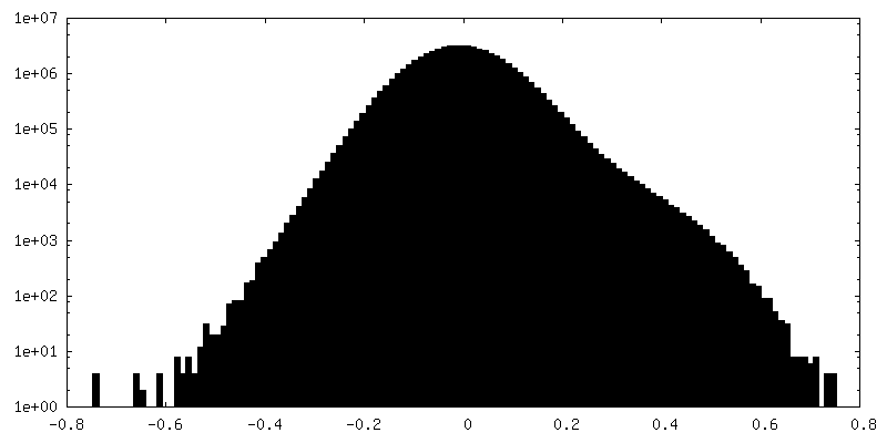
| Density Histograms |


- Sample components
-Entire : DpHF7
| Entire | Name: DpHF7 |
|---|---|
| Components |
|
-Supramolecule #1: DpHF7
| Supramolecule | Name: DpHF7 / type: complex / ID: 1 / Parent: 0 / Macromolecule list: all |
|---|---|
| Source (natural) | Organism: synthetic construct (others) |
-Macromolecule #1: DpHF7 filament
| Macromolecule | Name: DpHF7 filament / type: protein_or_peptide / ID: 1 / Number of copies: 20 / Enantiomer: LEVO |
|---|---|
| Source (natural) | Organism: synthetic construct (others) |
| Molecular weight | Theoretical: 26.640637 KDa |
| Recombinant expression | Organism:  Escherichia coli (E. coli) Escherichia coli (E. coli) |
| Sequence | String: MGSEVEILKA LLELKKSTAE LKRATASLRA ITEELKKNPS EDALVEHNRA IVEHNAIIVE NNRIIAAVLM LIVVAVGMTQ EIKKALEEL VASTAELKRA TASLRAITEE LKKNPSEDAL VEHNRAIVEH NAIIVENNRI IAAVLELIVR ALNLTDAEVI K ALIELRLS ...String: MGSEVEILKA LLELKKSTAE LKRATASLRA ITEELKKNPS EDALVEHNRA IVEHNAIIVE NNRIIAAVLM LIVVAVGMTQ EIKKALEEL VASTAELKRA TASLRAITEE LKKNPSEDAL VEHNRAIVEH NAIIVENNRI IAAVLELIVR ALNLTDAEVI K ALIELRLS TLELVAATAS LREITEELKK NPSEDALVEH NRAIVEHNAI IVENNRIIAA VLELIVGGSG GSGGSGGSSL EH HHHHH |
-Experimental details
-Structure determination
| Method | cryo EM |
|---|---|
 Processing Processing | helical reconstruction |
| Aggregation state | filament |
-Sample preparation
| Buffer | pH: 8 |
|---|---|
| Vitrification | Cryogen name: ETHANE |
- Electron microscopy
| Microscope | FEI TITAN KRIOS |
|---|---|
| Electron beam | Acceleration voltage: 300 kV / Electron source: FIELD EMISSION GUN |
| Electron optics | Illumination mode: FLOOD BEAM / Imaging mode: BRIGHT FIELDBright-field microscopy / Nominal defocus max: 2.0 µm / Nominal defocus min: 0.5 µm |
| Image recording | Film or detector model: GATAN K2 SUMMIT (4k x 4k) / Detector mode: SUPER-RESOLUTION / Average electron dose: 90.0 e/Å2 |
| Experimental equipment |  Model: Titan Krios / Image courtesy: FEI Company |
-Image processing
| Startup model | Type of model: OTHER |
|---|---|
| Final angle assignment | Type: NOT APPLICABLE / Software - Name: cryoSPARC |
| Final reconstruction | Applied symmetry - Helical parameters - Δz: 24.0 Å Applied symmetry - Helical parameters - Δ&Phi: -43.1 ° Applied symmetry - Helical parameters - Axial symmetry: D2 (2x2 fold dihedral )Resolution.type: BY AUTHOR / Resolution: 3.3 Å / Resolution method: FSC 0.143 CUT-OFF / Software - Name: cryoSPARC / Number images used: 56749 |
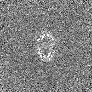
| FSC plot (resolution estimation) | 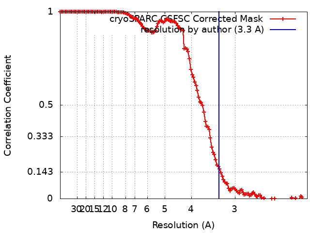 |
-Atomic model buiding 1
| Initial model | Chain - Initial model type: in silico model |
|---|---|
| Output model | PDB-8ub3: |
