 Movie
Movie Controller


+ Open data

- Basic information
| Entry | 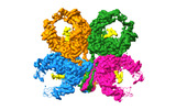 | |||||||||
|---|---|---|---|---|---|---|---|---|---|---|
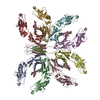
| Title | Cryo-EM structure of fish immunogloblin M-Fc | |||||||||
 Map data Map data | ||||||||||
 Sample Sample |
| |||||||||
 Keywords Keywords | Immunoglobulin M / IgM /  immune system immune system | |||||||||
| Biological species |  Oncorhynchus mykiss (rainbow trout) Oncorhynchus mykiss (rainbow trout) | |||||||||
| Method | single particle reconstruction / cryo EM / Resolution: 2.78 Å | |||||||||
 Authors Authors | Lyu M / Stadtmueller BM / Malyutin AG | |||||||||
| Funding support |  United States, 1 items United States, 1 items
| |||||||||
 Citation Citation | Journal: Nat Commun / Year: 2023 Title: The structure of the teleost Immunoglobulin M core provides insights on polymeric antibody evolution, assembly, and function. Authors: Mengfan Lyu / Andrey G Malyutin / Beth M Stadtmueller / Abstract: Polymeric (p) immunoglobulins (Igs) serve broad functions during vertebrate immune responses. Typically, pIgs contain between two and six Ig monomers, each with two antigen binding fragments and one ...Polymeric (p) immunoglobulins (Igs) serve broad functions during vertebrate immune responses. Typically, pIgs contain between two and six Ig monomers, each with two antigen binding fragments and one fragment crystallization (Fc). In addition, many pIgs assemble with a joining-chain (JC); however, the number of monomers and potential to include JC vary with species and heavy chain class. Here, we report the cryo-electron microscopy structure of IgM from a teleost (t) species, which does not encode JC. The structure reveals four tIgM Fcs linked through eight C-terminal tailpieces (Tps), which adopt a single β-sandwich-like domain (Tp assembly) located between two Fcs. Specifically, two of eight heavy chains fold uniquely, resulting in a structure distinct from mammalian IgM, which typically contains five IgM monomers, one JC and a centrally-located Tp assembly. Together with mutational analysis, structural data indicate that pIgs have evolved a range of assembly mechanisms and structures, each likely to support unique antibody effector functions. | |||||||||
| History |
|
- Structure visualization
| Supplemental images |
|---|

- Downloads & links
-EMDB archive
| Map data | emd_40054.map.gz | 83.3 MB |  EMDB map data format EMDB map data format | |
|---|---|---|---|---|
| Header (meta data) | emd-40054-v30.xmlemd-40054.xml | 17.3 KB 17.3 KB | Display Display | EMDB header |
| FSC (resolution estimation) | emd_40054_fsc.xml | 11.6 KB | Display | FSC data file |
| Images |  emd_40054.png emd_40054.png | 84.8 KB | ||
| Filedesc metadata | emd-40054.cif.gz | 6.3 KB | ||
| Others | emd_40054_half_map_1.map.gzemd_40054_half_map_2.map.gz | 154.3 MB 154.3 MB | ||
| Archive directory |  http://ftp.pdbj.org/pub/emdb/structures/EMD-40054ftp://ftp.pdbj.org/pub/emdb/structures/EMD-40054 http://ftp.pdbj.org/pub/emdb/structures/EMD-40054ftp://ftp.pdbj.org/pub/emdb/structures/EMD-40054 | HTTPS FTP |
-Related structure data
-Links
| EMDB pages | EMDB (EBI/PDBe) / EMDataResource |
|---|
-Map
| File | Download / File: emd_40054.map.gz / Format: CCP4 / Size: 166.4 MB / Type: IMAGE STORED AS FLOATING POINT NUMBER (4 BYTES) | ||||||||||||||||||||
|---|---|---|---|---|---|---|---|---|---|---|---|---|---|---|---|---|---|---|---|---|---|
| Voxel size | X=Y=Z: 0.855 Å | ||||||||||||||||||||
| Density |
| ||||||||||||||||||||
| Symmetry | Space group: 1 | ||||||||||||||||||||
| Details | EMDB XML:
|
-Supplemental data
-Half map: #2
| File | emd_40054_half_map_1.map | ||||||||||||
|---|---|---|---|---|---|---|---|---|---|---|---|---|---|
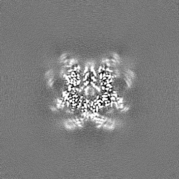
| Projections & Slices |
| ||||||||||||
| Density Histograms |
 Z
Z Y
Y X
X


-Half map: #1
| File | emd_40054_half_map_2.map | ||||||||||||
|---|---|---|---|---|---|---|---|---|---|---|---|---|---|
| Projections & Slices |
| ||||||||||||
| Density Histograms |


- Sample components
-Entire : rainbow trout (Oncorhynchus mykiss) immunoglobulin M Fc
| Entire | Name: rainbow trout (Oncorhynchus mykiss) immunoglobulin M Fc |
|---|---|
| Components |
|
-Supramolecule #1: rainbow trout (Oncorhynchus mykiss) immunoglobulin M Fc
| Supramolecule | Name: rainbow trout (Oncorhynchus mykiss) immunoglobulin M Fc type: complex / ID: 1 / Parent: 0 / Macromolecule list: all Details: Heavy chain constant domains 3 and 4 including C-terminal tailpiece |
|---|---|
| Source (natural) | Organism: Oncorhynchus mykiss (rainbow trout) |
-Macromolecule #1: Teleost immunoglobulin M protein
| Macromolecule | Name: Teleost immunoglobulin M protein / type: protein_or_peptide / ID: 1 / Number of copies: 8 / Enantiomer: LEVO |
|---|---|
| Source (natural) | Organism: Oncorhynchus mykiss (rainbow trout) |
| Molecular weight | Theoretical: 26.998514 KDa |
| Recombinant expression | Organism:  Homo sapiens (human) Homo sapiens (human) |
| Sequence | String: HHHHHHHGHL VVITIIEPSL EDMLMNKKAQ LVCDVNELVP GFLSVKWEND NGKTLTSRKG VTDKIAILDI TYEDWSNGTV FYCAVDHME NLGDLVKKAY KRETGGVPQR PSVFLLAPAE QTSDNTVTLT CYVKDFYPKD VLVAWLVDDE PVERTSSSAL Y QFNTTSQI ...String: HHHHHHHGHL VVITIIEPSL EDMLMNKKAQ LVCDVNELVP GFLSVKWEND NGKTLTSRKG VTDKIAILDI TYEDWSNGTV FYCAVDHME NLGDLVKKAY KRETGGVPQR PSVFLLAPAE QTSDNTVTLT CYVKDFYPKD VLVAWLVDDE PVERTSSSAL Y QFNTTSQI QSGRTYSVYS QLTFSNDLWK NEEVVYSCVV YHESMIKSTN IIMRTIDRTS NQPNLVNLSL NVPQRCMAQ |
-Experimental details
-Structure determination
| Method | cryo EM |
|---|---|
 Processing Processing | single particle reconstruction |
| Aggregation state | particle |
-Sample preparation
| Concentration | 0.1 mg/mL | |||||||||
|---|---|---|---|---|---|---|---|---|---|---|
| Buffer | pH: 7.8 Component:
| |||||||||
| Grid | Model: Quantifoil R1.2/1.3 / Material: GOLD / Mesh: 300 / Support film - Material: GOLD / Support film - topology: HOLEY / Pretreatment - Type: GLOW DISCHARGE / Pretreatment - Time: 90 sec. / Pretreatment - Atmosphere: AIR / Pretreatment - Pressure: 0.029 kPa / Details: Pelco easiGlow at 25 mA | |||||||||
| Vitrification | Cryogen name: ETHANE / Chamber humidity: 100 % / Chamber temperature: 277.15 K / Instrument: FEI VITROBOT MARK IV |
- Electron microscopy
| Microscope | FEI TITAN KRIOS |
|---|---|
| Electron beam | Acceleration voltage: 300 kV / Electron source: FIELD EMISSION GUN |
| Electron optics | C2 aperture diameter: 50.0 µm / Illumination mode: FLOOD BEAM / Imaging mode: BRIGHT FIELDBright-field microscopy / Cs: 2.7 mm / Nominal defocus max: 3.0 µm / Nominal defocus min: 0.6 µm / Nominal magnification: 105000 |
| Specialist optics | Energy filter - Name: GIF Bioquantum / Energy filter - Slit width: 20 eV |
| Sample stage | Specimen holder model: FEI TITAN KRIOS AUTOGRID HOLDER / Cooling holder cryogen: NITROGEN |
| Image recording | Film or detector model: GATAN K3 BIOQUANTUM (6k x 4k) / Digitization - Dimensions - Width: 5760 pixel / Digitization - Dimensions - Height: 4092 pixel / Number grids imaged: 1 / Number real images: 3618 / Average electron dose: 60.0 e/Å2 Details: Images were collected with SerialEM using beam image-shift in a 3x3x3 pattern. A total of ~60 e/A2 was fractionated into 40 frames movies. |
| Experimental equipment |  Model: Titan Krios / Image courtesy: FEI Company |
-Image processing
| Particle selection | Number selected: 1300000 |
|---|---|
| Startup model | Type of model: NONE |
| Initial angle assignment | Type: RANDOM ASSIGNMENT / Software - Name: cryoSPARC (ver. 4.0) |
| Final 3D classification | Software - Name: cryoSPARC (ver. 4.0) |
| Final angle assignment | Type: MAXIMUM LIKELIHOOD / Software - Name: cryoSPARC (ver. 4.0) |
| Final reconstruction | Applied symmetry - Point group: C2 (2 fold cyclic) / Resolution.type: BY AUTHOR / Resolution: 2.78 Å / Resolution method: FSC 0.143 CUT-OFF / Software - Name: cryoSPARC (ver. 4.0) / Number images used: 61000 |
| FSC plot (resolution estimation) | 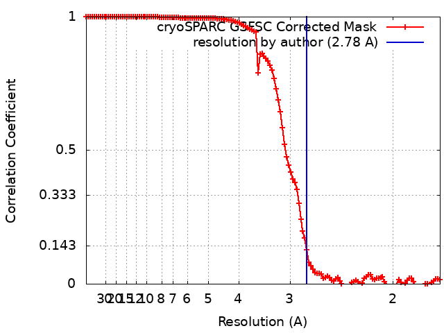 |
