 Movie
Movie Controller


+ Open data

- Basic information
| Entry |  | ||||||||||||
|---|---|---|---|---|---|---|---|---|---|---|---|---|---|
| Title | Cryo-EM structure of the hamster prion 23-144 fibril at pH 3.7 | ||||||||||||
 Map data Map data | |||||||||||||
 Sample Sample |
| ||||||||||||
 Keywords Keywords |  prion / 23-144 / hamster / PrP / PROTEIN FIBRIL prion / 23-144 / hamster / PrP / PROTEIN FIBRIL | ||||||||||||
| Function / homology |  Function and homology information Function and homology informationregulation of glutamate receptor signaling pathway / regulation of calcium ion import across plasma membrane / aspartic-type endopeptidase inhibitor activity / glycosaminoglycan binding / negative regulation of interleukin-17 production / regulation of potassium ion transmembrane transport / negative regulation of dendritic spine maintenance / type 5 metabotropic glutamate receptor binding / cupric ion binding / negative regulation of calcineurin-NFAT signaling cascade ...regulation of glutamate receptor signaling pathway / regulation of calcium ion import across plasma membrane / aspartic-type endopeptidase inhibitor activity / glycosaminoglycan binding / negative regulation of interleukin-17 production / regulation of potassium ion transmembrane transport / negative regulation of dendritic spine maintenance / type 5 metabotropic glutamate receptor binding / cupric ion binding / negative regulation of calcineurin-NFAT signaling cascade / negative regulation of interleukin-2 production / negative regulation of T cell receptor signaling pathway / cuprous ion binding / negative regulation of amyloid-beta formation / negative regulation of activated T cell proliferation / : / negative regulation of type II interferon production / positive regulation of protein targeting to membrane / side of membrane / inclusion body / cellular response to copper ion / neuron projection maintenance / molecular condensate scaffold activity / protein sequestering activity / negative regulation of protein phosphorylation / molecular function activator activity / positive regulation of protein localization to plasma membrane / protein destabilization / protein homooligomerization / terminal bouton / cellular response to amyloid-beta / positive regulation of neuron apoptotic process / positive regulation of peptidyl-tyrosine phosphorylation / cellular response to xenobiotic stimulus / signaling receptor activity / amyloid-beta binding / microtubule binding / nuclear membrane / response to oxidative stress / protease binding / amyloid fibril formation / learning or memory / regulation of cell cycle / cell cycle / membrane raft / copper ion binding / dendrite / protein-containing complex binding / negative regulation of apoptotic process / Golgi apparatus / cell surface / endoplasmic reticulum / identical protein binding / plasma membrane / cytosolSimilarity search - Function | ||||||||||||
| Biological species |  Mesocricetus auratus (golden hamster) Mesocricetus auratus (golden hamster) | ||||||||||||
| Method | helical reconstruction / cryo EM / Resolution: 2.88 Å | ||||||||||||
 Authors Authors | Lee C-H / Saw J-E / Chen E / Wang C-H / Chen R | ||||||||||||
| Funding support |  Taiwan, 3 items Taiwan, 3 items
| ||||||||||||
 Citation Citation | Journal: J Mol Biol / Year: 2024 Title: The Positively Charged Cluster in the N-terminal Disordered Region may Affect Prion Protein Misfolding: Cryo-EM Structure of Hamster PrP(23-144) Fibrils. Authors: Chih-Hsuan Lee / Jing-Ee Saw / Eric H-L Chen / Chun-Hsiung Wang / Takayuki Uchihashi / Rita P-Y Chen /  Abstract: Prions, the misfolding form of prion proteins, are contagious proteinaceous macromolecules. Recent studies have shown that infectious prion fibrils formed in the brain and non-infectious fibrils ...Prions, the misfolding form of prion proteins, are contagious proteinaceous macromolecules. Recent studies have shown that infectious prion fibrils formed in the brain and non-infectious fibrils formed from recombinant prion protein in a partially denaturing condition have distinct structures. The amyloid core of the in vitro-prepared non-infectious fibrils starts at about residue 160, while that of infectious prion fibrils formed in the brain involves a longer sequence (residues ∼90-230) of structural conversion. The C-terminal truncated prion protein PrP(23-144) can form infectious fibrils under certain conditions and cause disease in animals. In this study, we used cryogenic electron microscopy (cryo-EM) to resolve the structure of hamster sHaPrP(23-144) fibrils prepared at pH 3.7. This 2.88 Å cryo-EM structure has an amyloid core covering residues 94-144. It comprises two protofilaments, each containing five β-strands arranged as a long hairpin plus an N-terminal β-strand. This N-terminal β-strand resides in a positively charged cluster region (named PCC2; sequence 96-111), which interacts with the turn region of the opposite protofilaments' hairpin to stabilize the fibril structure. Interestingly, this sHaPrP(23-144) fibril structure differs from a recently reported structure formed by the human or mouse counterpart at pH 6.5. Moreover, sHaPrP(23-144) fibrils have many structural features in common with infectious prions. Whether this structure is infectious remains to be determined. More importantly, the sHaPrP(23-144) structure is different from the sHaPrP(108-144) fibrils prepared in the same fibrillization buffer, indicating that the N-terminal disordered region, possibly the positively charged cluster, influences the misfolding pathway of the prion protein. | ||||||||||||
| History |
|
- Structure visualization
| Supplemental images |
|---|

- Downloads & links
-EMDB archive
| Map data | emd_37963.map.gz | 9.2 MB | EMDB map data format | |
|---|---|---|---|---|
| Header (meta data) | emd-37963-v30.xmlemd-37963.xml | 14.3 KB 14.3 KB | Display Display | EMDB header |
| FSC (resolution estimation) | emd_37963_fsc.xml | 10.3 KB | Display | FSC data file |
| Images |  emd_37963.png emd_37963.png | 59.1 KB | ||
| Filedesc metadata | emd-37963.cif.gz | 5.1 KB | ||
| Others | emd_37963_half_map_1.map.gzemd_37963_half_map_2.map.gz | 71.4 MB 71.3 MB | ||
| Archive directory |  http://ftp.pdbj.org/pub/emdb/structures/EMD-37963ftp://ftp.pdbj.org/pub/emdb/structures/EMD-37963 http://ftp.pdbj.org/pub/emdb/structures/EMD-37963ftp://ftp.pdbj.org/pub/emdb/structures/EMD-37963 | HTTPS FTP |
-Related structure data
| Related structure data | 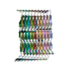 8wzxMC M: atomic model generated by this map C: citing same article ( |
|---|---|
| Similar structure data |
-Links
| EMDB pages | EMDB (EBI/PDBe) / EMDataResource |
|---|---|
| Related items in Molecule of the Month |


-Map
| File | Download / File: emd_37963.map.gz / Format: CCP4 / Size: 91.1 MB / Type: IMAGE STORED AS FLOATING POINT NUMBER (4 BYTES) | ||||||||||||||||||||
|---|---|---|---|---|---|---|---|---|---|---|---|---|---|---|---|---|---|---|---|---|---|
| Voxel size | X=Y=Z: 0.83 Å | ||||||||||||||||||||
| Density |
| ||||||||||||||||||||
| Symmetry | Space group: 1 | ||||||||||||||||||||
| Details | EMDB XML:
|
-Supplemental data
-Half map: #1
| File | emd_37963_half_map_1.map | ||||||||||||
|---|---|---|---|---|---|---|---|---|---|---|---|---|---|
| Projections & Slices |
| ||||||||||||
| Density Histograms |
 Z
Z Y
Y X
X


-Half map: #2
| File | emd_37963_half_map_2.map | ||||||||||||
|---|---|---|---|---|---|---|---|---|---|---|---|---|---|
| Projections & Slices |
| ||||||||||||
| Density Histograms |


- Sample components
-Entire : hamster prion 23-144 fibril
| Entire | Name: hamster prion 23-144 fibril |
|---|---|
| Components |
|
-Supramolecule #1: hamster prion 23-144 fibril
| Supramolecule | Name: hamster prion 23-144 fibril / type: complex / ID: 1 / Parent: 0 / Macromolecule list: all |
|---|---|
| Source (natural) | Organism: Mesocricetus auratus (golden hamster) |
-Macromolecule #1: Major prion protein
| Macromolecule | Name: Major prion protein / type: protein_or_peptide / ID: 1 / Number of copies: 20 / Enantiomer: LEVO |
|---|---|
| Source (natural) | Organism: Mesocricetus auratus (golden hamster) |
| Molecular weight | Theoretical: 12.376642 KDa |
| Recombinant expression | Organism:  Escherichia coli BL21(DE3) (bacteria) Escherichia coli BL21(DE3) (bacteria) |
| Sequence | String: GKKRPKPGGW NTGGSRYPGQ GSPGGNRYPP QGGGTWGQPH GGGWGQPHGG GWGQPHGGGW GQPHGGGWGQ GGGTHNQWNK PSKPKTNMK HMAGAAAAGA VVGGLGGYML GSAMSRPMMH FGND UniProtKB: Major prion protein |
-Experimental details
-Structure determination
| Method | cryo EM |
|---|---|
 Processing Processing | helical reconstruction |
| Aggregation state | filament |
-Sample preparation
| Buffer | pH: 3.7 / Details: 20 mM NaOAc, 140 mM NaCl |
|---|---|
| Vitrification | Cryogen name: ETHANE |
- Electron microscopy
| Microscope | TFS KRIOS |
|---|---|
| Electron beam | Acceleration voltage: 300 kV / Electron source: FIELD EMISSION GUN |
| Electron optics | Illumination mode: FLOOD BEAM / Imaging mode: BRIGHT FIELDBright-field microscopy / Nominal defocus max: 2.0 µm / Nominal defocus min: 1.4000000000000001 µm |
| Image recording | Film or detector model: GATAN K3 BIOQUANTUM (6k x 4k) / Average electron dose: 50.0 e/Å2 |
| Experimental equipment |  Model: Titan Krios / Image courtesy: FEI Company |
-Image processing
| Startup model | Type of model: INSILICO MODEL |
|---|---|
| Final angle assignment | Type: NOT APPLICABLE |
| Final reconstruction | Applied symmetry - Helical parameters - Δz: 2.38 Å Applied symmetry - Helical parameters - Δ&Phi: 179.217 ° Applied symmetry - Helical parameters - Axial symmetry: C1 (asymmetric) Resolution.type: BY AUTHOR / Resolution: 2.88 Å / Resolution method: FSC 0.143 CUT-OFF / Software - Name: RELION / Number images used: 288368 |
| FSC plot (resolution estimation) |  |
-Atomic model buiding 1
| Refinement | Space: REAL / Protocol: AB INITIO MODEL / Overall B value: 101.7 |
|---|---|
| Output model | PDB-8wzx: |
