 Movie
Movie Controller


+ Open data

- Basic information
| Entry |  | |||||||||
|---|---|---|---|---|---|---|---|---|---|---|
| Title | structure of AtHKT1;1 in KCl at 2.8 Angstroms resolution | |||||||||
 Map data Map data | ||||||||||
 Sample Sample |
| |||||||||
 Keywords Keywords | HKT / ion selectivity /  salt tolerance / TRANSPORT PROTEIN salt tolerance / TRANSPORT PROTEIN | |||||||||
| Function / homology |  Function and homology information Function and homology informationpotassium ion transmembrane transporter activity / response to osmotic stress / sodium ion transport / response to salt stress / potassium ion transport / plasma membraneSimilarity search - Function | |||||||||
| Biological species |  Arabidopsis thaliana (thale cress) Arabidopsis thaliana (thale cress) | |||||||||
| Method | single particle reconstruction / cryo EM / Resolution: 2.8 Å | |||||||||
 Authors Authors | Wang JQ | |||||||||
| Funding support |  China, 1 items China, 1 items
| |||||||||
 Citation Citation | Journal: To Be Published Title: structure of AtHKT1;1 in KCl at 2.8 Angstroms resolution Authors: Wang JQ | |||||||||
| History |
|
- Structure visualization
| Supplemental images |
|---|

- Downloads & links
-EMDB archive
| Map data | emd_37377.map.gz | 28.7 MB | EMDB map data format | |
|---|---|---|---|---|
| Header (meta data) | emd-37377-v30.xmlemd-37377.xml | 12.9 KB 12.9 KB | Display Display | EMDB header |
| Images | 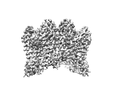 emd_37377.png emd_37377.png | 65.3 KB | ||
| Filedesc metadata | emd-37377.cif.gz | 5.2 KB | ||
| Others | emd_37377_half_map_1.map.gzemd_37377_half_map_2.map.gz | 28 MB 28.1 MB | ||
| Archive directory |  http://ftp.pdbj.org/pub/emdb/structures/EMD-37377ftp://ftp.pdbj.org/pub/emdb/structures/EMD-37377 http://ftp.pdbj.org/pub/emdb/structures/EMD-37377ftp://ftp.pdbj.org/pub/emdb/structures/EMD-37377 | HTTPS FTP |
-Related structure data
| Related structure data | 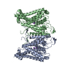 8w9oMC M: atomic model generated by this map C: citing same article ( |
|---|---|
| Similar structure data |
-Links
| EMDB pages | EMDB (EBI/PDBe) / EMDataResource |
|---|
-Map
| File | Download / File: emd_37377.map.gz / Format: CCP4 / Size: 35.3 MB / Type: IMAGE STORED AS FLOATING POINT NUMBER (4 BYTES) | ||||||||||||||||||||
|---|---|---|---|---|---|---|---|---|---|---|---|---|---|---|---|---|---|---|---|---|---|
| Voxel size | X=Y=Z: 0.93 Å | ||||||||||||||||||||
| Density |
| ||||||||||||||||||||
| Symmetry | Space group: 1 | ||||||||||||||||||||
| Details | EMDB XML:
|
-Supplemental data
-Half map: #1
| File | emd_37377_half_map_1.map | ||||||||||||
|---|---|---|---|---|---|---|---|---|---|---|---|---|---|
| Projections & Slices |
| ||||||||||||
| Density Histograms |
 Z
Z Y
Y X
X


-Half map: #2
| File | emd_37377_half_map_2.map | ||||||||||||
|---|---|---|---|---|---|---|---|---|---|---|---|---|---|
| Projections & Slices |
| ||||||||||||
| Density Histograms |


- Sample components
-Entire : structure of AtHKT1;1 in KCl at 2.8 Angstroms resolution
| Entire | Name: structure of AtHKT1;1 in KCl at 2.8 Angstroms resolution |
|---|---|
| Components |
|
-Supramolecule #1: structure of AtHKT1;1 in KCl at 2.8 Angstroms resolution
| Supramolecule | Name: structure of AtHKT1;1 in KCl at 2.8 Angstroms resolution type: complex / ID: 1 / Parent: 0 / Macromolecule list: #1 |
|---|---|
| Source (natural) | Organism: Arabidopsis thaliana (thale cress) |
-Macromolecule #1: Sodium transporter HKT1
| Macromolecule | Name: Sodium transporter HKT1 / type: protein_or_peptide / ID: 1 / Number of copies: 2 / Enantiomer: LEVO |
|---|---|
| Source (natural) | Organism: Arabidopsis thaliana (thale cress) |
| Molecular weight | Theoretical: 57.504965 KDa |
| Recombinant expression | Organism:  Homo sapiens (human) Homo sapiens (human) |
| Sequence | String: MDRVVAKIAK IRSQLTKLRS LFFLYFIYFL FFSFLGFLAL KITKPRTTSR PHDFDLFFTS VSAITVSSMS TVDMEVFSNT QLIFLTILM FLGGEIFTSF LNLYVSYFTK FVFPHNKIRH ILGSYNSDSS IEDRCDVETV TDYREGLIKI DERASKCLYS V VLSYHLVT ...String: MDRVVAKIAK IRSQLTKLRS LFFLYFIYFL FFSFLGFLAL KITKPRTTSR PHDFDLFFTS VSAITVSSMS TVDMEVFSNT QLIFLTILM FLGGEIFTSF LNLYVSYFTK FVFPHNKIRH ILGSYNSDSS IEDRCDVETV TDYREGLIKI DERASKCLYS V VLSYHLVT NLVGSVLLLV YVNFVKTARD VLSSKEISPL TFSVFTTVST FANCGFVPTN ENMIIFRKNS GLIWLLIPQV LM GNTLFPC FLVLLIWGLY KITKRDEYGY ILKNHNKMGY SHLLSVRLCV LLGVTVLGFL IIQLLFFCAF EWTSESLEGM SSY EKLVGS LFQVVNSRHT GETIVDLSTL SPAILVLFIL MMYLPPYTLF MPLTEQKTIE KEGGDDDSEN GKKVKKSGLI VSQL SFLTI CIFLISITER QNLQRDPINF NVLNITLEVI SAYGNVGFTT GYSCERRVDI SDGGCKDASY GFAGRWSPMG KFVLI IVMF YGRFKQFTAK SGRAWILYPS SS UniProtKB: Sodium transporter HKT1 |
-Macromolecule #2: POTASSIUM ION
| Macromolecule | Name: POTASSIUM ION / type: ligand / ID: 2 / Number of copies: 4 / Formula: K |
|---|---|
| Molecular weight | Theoretical: 39.098 Da |
-Experimental details
-Structure determination
| Method | cryo EM |
|---|---|
 Processing Processing | single particle reconstruction |
| Aggregation state | particle |
-Sample preparation
| Buffer | pH: 7.5 |
|---|---|
| Vitrification | Cryogen name: ETHANE |
- Electron microscopy
| Microscope | FEI TITAN KRIOS |
|---|---|
| Electron beam | Acceleration voltage: 300 kV / Electron source: FIELD EMISSION GUN |
| Electron optics | Illumination mode: FLOOD BEAM / Imaging mode: BRIGHT FIELDBright-field microscopy / Cs: 2.7 mm / Nominal defocus max: 1.6 µm / Nominal defocus min: 0.8 µm |
| Image recording | Film or detector model: GATAN K2 QUANTUM (4k x 4k) / Average exposure time: 8.0 sec. / Average electron dose: 52.0 e/Å2 |
| Experimental equipment |  Model: Titan Krios / Image courtesy: FEI Company |
-Image processing
| Startup model | Type of model: OTHER |
|---|---|
| Initial angle assignment | Type: MAXIMUM LIKELIHOOD |
| Final angle assignment | Type: MAXIMUM LIKELIHOOD |
| Final reconstruction | Resolution.type: BY AUTHOR / Resolution: 2.8 Å / Resolution method: FSC 0.143 CUT-OFF / Number images used: 54018 |
