Movie
Movie Controller

[English] 日本語
 Yorodumi
- EMDB-33792: Structure of GluN1a-GluN2D NMDA receptor in complex with agonists... -
+ Open data

- Basic information
| Entry |  | ||||||||||||||||||
|---|---|---|---|---|---|---|---|---|---|---|---|---|---|---|---|---|---|---|---|
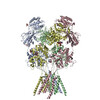
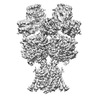
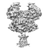
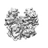
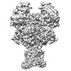
| Title | Structure of GluN1a-GluN2D NMDA receptor in complex with agonists glycine and glutamate. | ||||||||||||||||||
 Map data Map data | |||||||||||||||||||
 Sample Sample |
| ||||||||||||||||||
 Keywords Keywords |  ion channel / cryo-EM structure / glutamate receptor / synaptic protein / ELECTRON TRANSPORT ion channel / cryo-EM structure / glutamate receptor / synaptic protein / ELECTRON TRANSPORT | ||||||||||||||||||
| Function / homology |  Function and homology information Function and homology informationexcitatory chemical synaptic transmission / regulation of sensory perception of pain / Synaptic adhesion-like molecules / cellular response to L-glutamate / propylene metabolic process / response to glycine / voltage-gated monoatomic cation channel activity / glutamate-gated calcium ion channel activity / regulation of monoatomic cation transmembrane transport / Assembly and cell surface presentation of NMDA receptors ...excitatory chemical synaptic transmission / regulation of sensory perception of pain / Synaptic adhesion-like molecules / cellular response to L-glutamate / propylene metabolic process / response to glycine / voltage-gated monoatomic cation channel activity / glutamate-gated calcium ion channel activity / regulation of monoatomic cation transmembrane transport / Assembly and cell surface presentation of NMDA receptors / Neurexins and neuroligins / NMDA glutamate receptor activity / NMDA selective glutamate receptor complex / calcium ion transmembrane import into cytosol / protein heterotetramerization / glutamate binding / positive regulation of reactive oxygen species biosynthetic process / glycine binding / positive regulation of calcium ion transport into cytosol / Negative regulation of NMDA receptor-mediated neuronal transmission / startle response / Unblocking of NMDA receptors, glutamate binding and activation / regulation of neuronal synaptic plasticity / monoatomic cation transmembrane transport / positive regulation of excitatory postsynaptic potential / Long-term potentiation / monoatomic cation transport / ligand-gated monoatomic ion channel activity / excitatory synapse / calcium ion homeostasis / glutamate-gated receptor activity / synaptic cleft / presynaptic active zone membrane / ligand-gated monoatomic ion channel activity involved in regulation of presynaptic membrane potential / EPHB-mediated forward signaling / excitatory postsynaptic potential / hippocampal mossy fiber to CA3 synapse / regulation of membrane potential / ionotropic glutamate receptor signaling pathway / Ras activation upon Ca2+ influx through NMDA receptor / positive regulation of synaptic transmission, glutamatergic / adult locomotory behavior / transmitter-gated monoatomic ion channel activity involved in regulation of postsynaptic membrane potential / synaptic membrane / synaptic transmission, glutamatergic / long-term synaptic potentiation / postsynaptic density membrane / brain development / regulation of synaptic plasticity / visual learning / terminal bouton / synaptic vesicle / signaling receptor activity / amyloid-beta binding / chemical synaptic transmission / RAF/MAP kinase cascade / postsynaptic membrane / response to ethanol / dendritic spine / postsynaptic density / calmodulin binding / neuron projection / dendrite / synapse / glutamatergic synapse / calcium ion binding / protein-containing complex binding / endoplasmic reticulum membrane / cell surface / positive regulation of transcription by RNA polymerase II / plasma membrane / cytoplasmSimilarity search - Function | ||||||||||||||||||
| Biological species |  Homo sapiens (human) Homo sapiens (human) | ||||||||||||||||||
| Method | single particle reconstruction / cryo EM / Resolution: 3.9 Å | ||||||||||||||||||
 Authors Authors | Zhang JL / Zhu SJ / Zhang M | ||||||||||||||||||
| Funding support |  China, 5 items China, 5 items
| ||||||||||||||||||
 Citation Citation | Journal: Nat Struct Mol Biol / Year: 2023 Title: Distinct structure and gating mechanism in diverse NMDA receptors with GluN2C and GluN2D subunits. Authors: Jilin Zhang / Ming Zhang / Qinrui Wang / Han Wen / Zheyi Liu / Fangjun Wang / Yuhang Wang / Fenyong Yao / Nan Song / Zengwei Kou / Yang Li / Fei Guo / Shujia Zhu / Abstract: N-methyl-D-aspartate (NMDA) receptors are heterotetramers comprising two GluN1 and two alternate GluN2 (N2A-N2D) subunits. Here we report full-length cryo-EM structures of the human N1-N2D di- ...N-methyl-D-aspartate (NMDA) receptors are heterotetramers comprising two GluN1 and two alternate GluN2 (N2A-N2D) subunits. Here we report full-length cryo-EM structures of the human N1-N2D di-heterotetramer (di-receptor), rat N1-N2C di-receptor and N1-N2A-N2C tri-heterotetramer (tri-receptor) at a best resolution of 3.0 Å. The bilobate N-terminal domain (NTD) in N2D intrinsically adopts a closed conformation, leading to a compact NTD tetramer in the N1-N2D receptor. Additionally, crosslinking the ligand-binding domain (LBD) of two N1 protomers significantly elevated the channel open probability (Po) in N1-N2D di-receptors. Surprisingly, the N1-N2C di-receptor adopted both symmetric (minor) and asymmetric (major) conformations, the latter further locked by an allosteric potentiator, PYD-106, binding to a pocket between the NTD and LBD in only one N2C protomer. Finally, the N2A and N2C subunits in the N1-N2A-N2C tri-receptor display a conformation close to one protomer in the N1-N2A and N1-N2C di-receptors, respectively. These findings provide a comprehensive structural understanding of diverse function in major NMDA receptor subtypes. | ||||||||||||||||||
| History |
|
- Structure visualization
| Supplemental images |
|---|

- Downloads & links
-EMDB archive
| Map data | emd_33792.map.gz | 34.5 MB | EMDB map data format | |
|---|---|---|---|---|
| Header (meta data) | emd-33792-v30.xmlemd-33792.xml | 21.8 KB 21.8 KB | Display Display | EMDB header |
| FSC (resolution estimation) | emd_33792_fsc.xml | 9.3 KB | Display | FSC data file |
| Images | 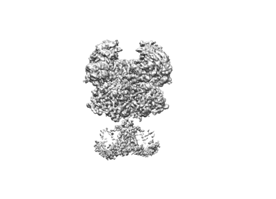 emd_33792.png emd_33792.png | 38.7 KB | ||
| Masks | emd_33792_msk_1.map | 67 MB | Mask map | |
| Others | emd_33792_half_map_1.map.gzemd_33792_half_map_2.map.gz | 61.8 MB 61.8 MB | ||
| Archive directory |  http://ftp.pdbj.org/pub/emdb/structures/EMD-33792ftp://ftp.pdbj.org/pub/emdb/structures/EMD-33792 http://ftp.pdbj.org/pub/emdb/structures/EMD-33792ftp://ftp.pdbj.org/pub/emdb/structures/EMD-33792 | HTTPS FTP |
-Related structure data
| Related structure data | 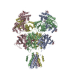 7yflMC 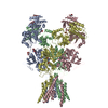 7yffC  7yfgC  7yfhC 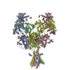 7yfiC 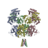 7yfmC  7yfoC  7yfrC  8hdkC C: citing same article ( M: atomic model generated by this map |
|---|---|
| Similar structure data |


-Links
| EMDB pages | EMDB (EBI/PDBe) / EMDataResource |
|---|---|
| Related items in Molecule of the Month |


-Map
| File | Download / File: emd_33792.map.gz / Format: CCP4 / Size: 67 MB / Type: IMAGE STORED AS FLOATING POINT NUMBER (4 BYTES) | ||||||||||||||||||||
|---|---|---|---|---|---|---|---|---|---|---|---|---|---|---|---|---|---|---|---|---|---|
| Voxel size | X=Y=Z: 1.06 Å | ||||||||||||||||||||
| Density |
| ||||||||||||||||||||
| Symmetry | Space group: 1 | ||||||||||||||||||||
| Details | EMDB XML:
|
-Supplemental data
-Mask #1
| File | emd_33792_msk_1.map | ||||||||||||
|---|---|---|---|---|---|---|---|---|---|---|---|---|---|
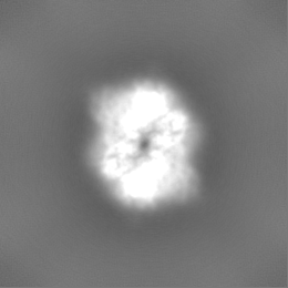
| Projections & Slices |
| ||||||||||||
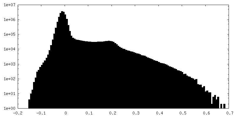
| Density Histograms |
 Z
Z Y
Y X
X


-Half map: #2
| File | emd_33792_half_map_1.map | ||||||||||||
|---|---|---|---|---|---|---|---|---|---|---|---|---|---|
| Projections & Slices |
| ||||||||||||
| Density Histograms |


-Half map: #1
| File | emd_33792_half_map_2.map | ||||||||||||
|---|---|---|---|---|---|---|---|---|---|---|---|---|---|
| Projections & Slices |
| ||||||||||||
| Density Histograms |


- Sample components
-Entire : NMDA receptor with NMDA 1 incorperated with NMDA 2D
| Entire | Name: NMDA receptor with NMDA 1 incorperated with NMDA 2D |
|---|---|
| Components |
|
-Supramolecule #1: NMDA receptor with NMDA 1 incorperated with NMDA 2D
| Supramolecule | Name: NMDA receptor with NMDA 1 incorperated with NMDA 2D / type: complex / ID: 1 / Parent: 0 / Macromolecule list: #1-#2 |
|---|---|
| Source (natural) | Organism: Homo sapiens (human) / Strain: Homo sapiens / Organ: brain / Tissue: brain / Organelle: synapse / Location in cell: plasma membrane |
| Molecular weight | Theoretical: 384.54 kDa/nm |
-Macromolecule #1: Glutamate receptor ionotropic, NMDA 1
| Macromolecule | Name: Glutamate receptor ionotropic, NMDA 1 / type: protein_or_peptide / ID: 1 / Number of copies: 2 / Enantiomer: LEVO |
|---|---|
| Source (natural) | Organism: Homo sapiens (human) |
| Molecular weight | Theoretical: 95.236078 KDa |
| Recombinant expression | Organism: Homo sapiens (human) |
| Sequence | String: MSTMRLLTLA LLFSCSVARA ACDPKIVNIG AVLSTRKHEQ MFREAVNQAN KRHGSWKIQL NATSVTHKPN AIQMALSVCE DLISSQVYA ILVSHPPTPN DHFTPTPVSY TAGFYRIPVL GLTTRMSIYS DKSIHLSFLR TVPPYSHQSS VWFEMMRVYS W NHIILLVS ...String: MSTMRLLTLA LLFSCSVARA ACDPKIVNIG AVLSTRKHEQ MFREAVNQAN KRHGSWKIQL NATSVTHKPN AIQMALSVCE DLISSQVYA ILVSHPPTPN DHFTPTPVSY TAGFYRIPVL GLTTRMSIYS DKSIHLSFLR TVPPYSHQSS VWFEMMRVYS W NHIILLVS DDHEGRAAQK RLETLLEERE SKAEKVLQFD PGTKNVTALL MEAKELEARV IILSASEDDA ATVYRAAAML NM TGSGYVW LVGEREISGN ALRYAPDGIL GLQLINGKNE SAHISDAVGV VAQAVHELLE KENITDPPRG CVGNTNIWKT GPL FKRVLM SSKYADGVTG RVEFNEDGDR KFANYSIMNL QNRKLVQVGI YNGTHVIPND RKIIWPGGET EKPRGYQMST RLKI VTIHQ EPFVYVKPTL SDGTCKEEFT VNGDPVKKVI CTGPNDTSPG SPRHTVPQCC YGFCIDLLIK LARTMNFTYE VHLVA DGKF GTQERVNNSN KKEWNGMMGE LLSGQADMIV APLTINNERA QYIEFSKPFK YQGLTILVKK EIPRSTLDSF MQPFQS TLW LLVGLSVHVV AVMLYLLDRF SPFGRFKVNS EEEEEDALTL SSAMWFSWGV LLNSGIGEGA PRSFSARILG MVWAGFA MI IVASYTANLA AFLVLDRPEE RITGINDPRL RNPSDKFIYA TVKQSSVDIY FRRQVELSTM YRHMEKHNYE SAAEAIQA V RDNKLHAFIW DSAVLEFEAS QKCDLVTTGE LFFRSGFGIG MRKDSPWKQN VSLSILKSHE NGFMEDLDKT WVRYQECDS RSNAPATLTF ENMAGVFMLV AGGIVAGIFL IFIEIAYKRH KDARRKQ UniProtKB: Glutamate receptor ionotropic, NMDA 1 |
-Macromolecule #2: Glutamate receptor ionotropic, NMDA 2D
| Macromolecule | Name: Glutamate receptor ionotropic, NMDA 2D / type: protein_or_peptide / ID: 2 / Number of copies: 2 / Enantiomer: LEVO |
|---|---|
| Source (natural) | Organism: Homo sapiens (human) |
| Molecular weight | Theoretical: 97.226414 KDa |
| Recombinant expression | Organism: Homo sapiens (human) |
| Sequence | String: MRGAGGPRGP RGPAKMLLLL ALACASPFPE EAPGPGGAGG PGGGLGGARP LNVALVFSGP AYAAEAARLG PAVAAAVRSP GLDVRPVAL VLNGSDPRSL VLQLCDLLSG LRVHGVVFED DSRAPAVAPI LDFLSAQTSL PIVAVHGGAA LVLTPKEKGS T FLQLGSST ...String: MRGAGGPRGP RGPAKMLLLL ALACASPFPE EAPGPGGAGG PGGGLGGARP LNVALVFSGP AYAAEAARLG PAVAAAVRSP GLDVRPVAL VLNGSDPRSL VLQLCDLLSG LRVHGVVFED DSRAPAVAPI LDFLSAQTSL PIVAVHGGAA LVLTPKEKGS T FLQLGSST EQQLQVIFEV LEEYDWTSFV AVTTRAPGHR AFLSYIEVLT DGSLVGWEHR GALTLDPGAG EAVLSAQLRS VS AQIRLLF CAREEAEPVF RAAEEAGLTG SGYVWFMVGP QLAGGGGSGA PGEPPLLPGG APLPAGLFAV RSAGWRDDLA RRV AAGVAV VARGAQALLR DYGFLPELGH DCRAQNRTHR GESLHRYFMN ITWDNRDYSF NEDGFLVNPS LVVISLTRDR TWEV VGSWE QQTLRLKYPL WSRYGRFLQP VDDTQHLTVA TLEERPFVIV EPADPISGTC IRDSVPCRSQ LNRTHSPPPD APRPE KRCC KGFCIDILKR LAHTIGFSYD LYLVTNGKHG KKIDGVWNGM IGEVFYQRAD MAIGSLTINE ERSEIVDFSV PFVETG ISV MVARSNGTVS PSAFLEPYSP AVWVMMFVMC LTVVAVTVFI FEYLSPVGYN RSLATGKRPG GSTFTIGKSI WLLWALV FN NSVPVENPRG TTSKIMVLVW AFFAVIFLAS YTANLAAFMI QEEYVDTVSG LSDRKFQRPQ EQYPPLKFGT VPNGSTEK N IRSNYPDMHS YMVRYNQPRV EEALTQLKAG KLDAFIYDAA VLNYMARKDE GCKLVTIGSG KVFATTGYGI ALHKGSRWK RPIDLALLQF LGDDEIEMLE RLWLSGICHN DKIEVMSSKL DIDNMAGVFY MLLVAMGLSL LVFAWEHLVY WRLRHCLGPA ASAWSHPQF EK UniProtKB: Glutamate receptor ionotropic, NMDA 2D |
-Macromolecule #4: GLYCINE
| Macromolecule | Name: GLYCINE / type: ligand / ID: 4 / Number of copies: 2 / Formula: GLY |
|---|---|
| Molecular weight | Theoretical: 75.067 Da |
| Chemical component information |  ChemComp-GLY: |
-Macromolecule #5: 2-acetamido-2-deoxy-beta-D-glucopyranose
| Macromolecule | Name: 2-acetamido-2-deoxy-beta-D-glucopyranose / type: ligand / ID: 5 / Number of copies: 10 / Formula: NAG |
|---|---|
| Molecular weight | Theoretical: 221.208 Da |
| Chemical component information |  ChemComp-NAG: |
-Macromolecule #6: GLUTAMIC ACID
| Macromolecule | Name: GLUTAMIC ACID / type: ligand / ID: 6 / Number of copies: 2 / Formula: GLU |
|---|---|
| Molecular weight | Theoretical: 147.129 Da |
| Chemical component information |  ChemComp-GLU: |
-Experimental details
-Structure determination
| Method | cryo EM |
|---|---|
 Processing Processing | single particle reconstruction |
| Aggregation state | particle |
-Sample preparation
| Concentration | 4.0 mg/mL |
|---|---|
| Buffer | pH: 8 |
| Grid | Model: Quantifoil R1.2/1.3 / Material: GOLD / Mesh: 300 / Pretreatment - Type: GLOW DISCHARGE / Pretreatment - Time: 60 sec. / Pretreatment - Atmosphere: AIR / Pretreatment - Pressure: 101.325 kPa |
| Vitrification | Cryogen name: ETHANE / Chamber humidity: 100 % / Chamber temperature: 281 K / Instrument: FEI VITROBOT MARK IV / Details: Blot for 3 seconds before plunging. |
| Details | This sample was monodisperse |
- Electron microscopy
| Microscope | FEI TITAN KRIOS |
|---|---|
| Electron beam | Acceleration voltage: 300 kV / Electron source: FIELD EMISSION GUN |
| Electron optics | Illumination mode: FLOOD BEAM / Imaging mode: BRIGHT FIELDBright-field microscopy / Nominal defocus max: 2.5 µm / Nominal defocus min: 1.5 µm |
| Image recording | Film or detector model: DIRECT ELECTRON DE-10 (5k x 4k) / Detector mode: SUPER-RESOLUTION / Digitization - Frames/image: 1-40 / Number grids imaged: 1 / Number real images: 3840 / Average electron dose: 60.0 e/Å2 |
| Experimental equipment |  Model: Titan Krios / Image courtesy: FEI Company |
-Image processing
| Particle selection | Number selected: 505204 |
|---|---|
| Startup model | Type of model: OTHER |
| Initial angle assignment | Type: OTHER / Software: (Name: RELION (ver. 3.1.1), cryoSPARC) |
| Final 3D classification | Number classes: 4 / Software - Name: cryoSPARC |
| Final angle assignment | Type: OTHER / Software - Name: cryoSPARC |
| Final reconstruction | Number classes used: 2 / Applied symmetry - Point group: C2 (2 fold cyclic) / Resolution.type: BY AUTHOR / Resolution: 3.9 Å / Resolution method: FSC 0.143 CUT-OFF / Software - Name: cryoSPARC / Number images used: 232194 |
| FSC plot (resolution estimation) |  |
-Atomic model buiding 1
| Initial model | PDB ID: Chain - Source name: PDB / Chain - Initial model type: experimental model |
|---|---|
| Refinement | Space: REAL / Protocol: FLEXIBLE FIT / Overall B value: 220 / Target criteria: Correlation coefficient |
| Output model | PDB-7yfl: |