[English] 日本語
Yorodumi
- EMDB-32920: S protein of SARS-CoV-2 in complex with 2G1 -

+
Open data


ID or keywords:

Loading...

-
Basic information

Entry
Database: EMDB / ID: EMD-32920
TitleS protein of SARS-CoV-2 in complex with 2G1
Map data
Sample
  • Complex: S protein of SARS-CoV-2 in complex with 2G1
    • Complex: S protein of SARS-CoV-2
      • Protein or peptide: Spike glycoproteinSpike protein
    • Complex: 2G1
      • Protein or peptide: heavy chain of 2G1
      • Protein or peptide: light chain of 2G1
  • Ligand: 2-acetamido-2-deoxy-beta-D-glucopyranose
  • Ligand: LINOLEIC ACID
KeywordsSARS-CoV-2 / VIRAL PROTEIN / VIRAL PROTEIN-IMMUNE SYSTEM complex
Function / homology
Function and homology information


Maturation of spike protein / viral translation / Translation of Structural Proteins / Virion Assembly and Release / host cell surface / host extracellular space / suppression by virus of host tetherin activity / Induction of Cell-Cell Fusion / structural constituent of virion / host cell endoplasmic reticulum-Golgi intermediate compartment membrane ...Maturation of spike protein / viral translation / Translation of Structural Proteins / Virion Assembly and Release / host cell surface / host extracellular space / suppression by virus of host tetherin activity / Induction of Cell-Cell Fusion / structural constituent of virion / host cell endoplasmic reticulum-Golgi intermediate compartment membrane / entry receptor-mediated virion attachment to host cell / receptor-mediated endocytosis of virus by host cell / Attachment and Entry / membrane fusion / positive regulation of viral entry into host cell / receptor-mediated virion attachment to host cell / receptor ligand activity / host cell surface receptor binding / fusion of virus membrane with host plasma membrane / fusion of virus membrane with host endosome membrane / viral envelope / virion attachment to host cell / SARS-CoV-2 activates/modulates innate and adaptive immune responses / host cell plasma membrane / virion membrane / membrane / identical protein binding / plasma membrane
Similarity search - Function
Spike (S) protein S1 subunit, receptor-binding domain, SARS-CoV-2 / Spike (S) protein S1 subunit, N-terminal domain, SARS-CoV-like / Betacoronavirus spike (S) glycoprotein S1 subunit N-terminal (NTD) domain profile. / Spike glycoprotein, N-terminal domain superfamily / Betacoronavirus spike (S) glycoprotein S1 subunit C-terminal (CTD) domain profile. / Spike glycoprotein, betacoronavirus / Spike (S) protein S1 subunit, receptor-binding domain, betacoronavirus / Spike S1 subunit, receptor binding domain superfamily, betacoronavirus / Betacoronavirus spike glycoprotein S1, receptor binding / Spike glycoprotein S1, N-terminal domain, betacoronavirus-like ...Spike (S) protein S1 subunit, receptor-binding domain, SARS-CoV-2 / Spike (S) protein S1 subunit, N-terminal domain, SARS-CoV-like / Betacoronavirus spike (S) glycoprotein S1 subunit N-terminal (NTD) domain profile. / Spike glycoprotein, N-terminal domain superfamily / Betacoronavirus spike (S) glycoprotein S1 subunit C-terminal (CTD) domain profile. / Spike glycoprotein, betacoronavirus / Spike (S) protein S1 subunit, receptor-binding domain, betacoronavirus / Spike S1 subunit, receptor binding domain superfamily, betacoronavirus / Betacoronavirus spike glycoprotein S1, receptor binding / Spike glycoprotein S1, N-terminal domain, betacoronavirus-like / Betacoronavirus-like spike glycoprotein S1, N-terminal / Spike glycoprotein S2, coronavirus, heptad repeat 1 / Spike glycoprotein S2, coronavirus, heptad repeat 2 / Coronavirus spike (S) glycoprotein S2 subunit heptad repeat 2 (HR2) region profile. / Coronavirus spike (S) glycoprotein S2 subunit heptad repeat 1 (HR1) region profile. / Spike glycoprotein S2 superfamily, coronavirus / Spike glycoprotein S2, coronavirus / Coronavirus spike glycoprotein S2 / Coronavirus spike glycoprotein S1, C-terminal / Coronavirus spike glycoprotein S1, C-terminal
Similarity search - Domain/homology
Biological speciesSevere acute respiratory syndrome coronavirus / Homo sapiens (human)
Methodsingle particle reconstruction / cryo EM / Resolution: 2.7 Å
AuthorsGuo YY / Zhang YY
Funding support China, 1 items
OrganizationGrant numberCountry
National Natural Science Foundation of China (NSFC)31971123 China
Citation
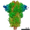
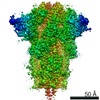
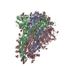
Journal: Cell Discov / Year: 2022
Title: Broad ultra-potent neutralization of SARS-CoV-2 variants by monoclonal antibodies specific to the tip of RBD.
Authors: Hang Ma / Yingying Guo / Haoneng Tang / Chien-Te K Tseng / Lei Wang / Huifang Zong / Zhenyu Wang / Yang He / Yunsong Chang / Shusheng Wang / Haiqiu Huang / Yong Ke / Yunsheng Yuan / Mingyuan ...Authors: Hang Ma / Yingying Guo / Haoneng Tang / Chien-Te K Tseng / Lei Wang / Huifang Zong / Zhenyu Wang / Yang He / Yunsong Chang / Shusheng Wang / Haiqiu Huang / Yong Ke / Yunsheng Yuan / Mingyuan Wu / Yuanyuan Zhang / Aleksandra Drelich / Kempaiah Rayavara Kempaiah / Bi-Hung Peng / Ailin Wang / Kaiyong Yang / Haiyang Yin / Junjun Liu / Yali Yue / Wenbo Xu / Shuangli Zhu / Tianjiao Ji / Xiaoju Zhang / Ziqi Wang / Gang Li / Guangchun Liu / Jingjing Song / Lingling Mu / ZongShang Xiang / Zhangyi Song / Hua Chen / Yanlin Bian / Baohong Zhang / Hui Chen / Jiawei Zhang / Yunji Liao / Li Zhang / Li Yang / Yi Chen / John Gilly / Xiaodong Xiao / Lei Han / Hua Jiang / Yueqing Xie / Qiang Zhou / Jianwei Zhu /
Abstract: Severe acute respiratory syndrome coronavirus 2 (SARS-CoV-2) variants of concern (VOCs) continue to wreak havoc across the globe. Higher transmissibility and immunologic resistance of VOCs bring ...Severe acute respiratory syndrome coronavirus 2 (SARS-CoV-2) variants of concern (VOCs) continue to wreak havoc across the globe. Higher transmissibility and immunologic resistance of VOCs bring unprecedented challenges to epidemic extinguishment. Here we describe a monoclonal antibody, 2G1, that neutralizes all current VOCs and has surprising tolerance to mutations adjacent to or within its interaction epitope. Cryo-electron microscopy structure showed that 2G1 bound to the tip of receptor binding domain (RBD) of spike protein with small contact interface but strong hydrophobic effect, which resulted in nanomolar to sub-nanomolar affinities to spike proteins. The epitope of 2G1 on RBD partially overlaps with angiotensin converting enzyme 2 (ACE2) interface, which enables 2G1 to block interaction between RBD and ACE2. The narrow binding epitope but high affinity bestow outstanding therapeutic efficacy upon 2G1 that neutralized VOCs with sub-nanomolar half maximal inhibitory concentration in vitro. In SARS-CoV-2, Beta or Delta variant-challenged transgenic mice and rhesus macaque models, 2G1 protected animals from clinical illness and eliminated viral burden, without serious impact to animal safety. Mutagenesis experiments suggest that 2G1 is potentially capable of dealing with emerging SARS-CoV-2 variants in the future. This report characterized the therapeutic antibodies specific to the tip of spike against SARS-CoV-2 variants and highlights the potential clinical applications as well as for developing vaccine and cocktail therapy.
History
DepositionFeb 21, 2022-
Header (metadata) releaseMar 9, 2022-
Map releaseMar 9, 2022-
UpdateNov 15, 2023-
Current statusNov 15, 2023Processing site: PDBj / Status: Released

-
Structure visualization

Movie
  • Surface view with section colored by density value
  • Surface level: 0.025
  • Imaged by UCSF Chimera
  • Download
  • Surface view colored by cylindrical radius
  • Surface level: 0.025
  • Imaged by UCSF Chimera
  • Download
  • Surface view with fitted model
  • Atomic models: PDB-7x08
  • Surface level: 0.025
  • Imaged by UCSF Chimera
  • Download
Movie viewer
Structure viewerEM map:
SurfViewMolmilJmol/JSmol
Supplemental images

Downloads & links

-
Map

FileDownload / File: emd_32920.map.gz / Format: CCP4 / Size: 91.1 MB / Type: IMAGE STORED AS FLOATING POINT NUMBER (4 BYTES)
Voxel sizeX=Y=Z: 1.087 Å
Density
Contour LevelBy AUTHOR: 0.025 / Movie #1: 0.025
Minimum - Maximum-0.11177208 - 0.224923
Average (Standard dev.)0.00003478838 (±0.0061582285)
SymmetrySpace group: 1
Details

EMDB XML:

Map geometry
Axis orderXYZ
Origin000
Dimensions288288288
Spacing288288288
CellA=B=C: 313.056 Å
α=β=γ: 90.0 °

CCP4 map header:

modeImage stored as Reals
Å/pix. X/Y/Z1.0871.0871.087
M x/y/z288288288
origin x/y/z0.0000.0000.000
length x/y/z313.056313.056313.056
α/β/γ90.00090.00090.000
start NX/NY/NZ000
NX/NY/NZ330330330
MAP C/R/S123
start NC/NR/NS000
NC/NR/NS288288288
D min/max/mean-0.1120.2250.000

-
Supplemental data

-
Sample components

-
Entire : S protein of SARS-CoV-2 in complex with 2G1

EntireName: S protein of SARS-CoV-2 in complex with 2G1
Components
  • Complex: S protein of SARS-CoV-2 in complex with 2G1
    • Complex: S protein of SARS-CoV-2
      • Protein or peptide: Spike glycoproteinSpike protein
    • Complex: 2G1
      • Protein or peptide: heavy chain of 2G1
      • Protein or peptide: light chain of 2G1
  • Ligand: 2-acetamido-2-deoxy-beta-D-glucopyranose
  • Ligand: LINOLEIC ACID

-
Supramolecule #1: S protein of SARS-CoV-2 in complex with 2G1

SupramoleculeName: S protein of SARS-CoV-2 in complex with 2G1 / type: complex / ID: 1 / Parent: 0 / Macromolecule list: #1-#3

-
Supramolecule #2: S protein of SARS-CoV-2

SupramoleculeName: S protein of SARS-CoV-2 / type: complex / ID: 2 / Parent: 1 / Macromolecule list: #1
Source (natural)Organism: Severe acute respiratory syndrome coronavirus

-
Supramolecule #3: 2G1

SupramoleculeName: 2G1 / type: complex / ID: 3 / Parent: 1 / Macromolecule list: #2-#3
Source (natural)Organism: Homo sapiens (human)

-
Macromolecule #1: Spike glycoprotein

MacromoleculeName: Spike glycoprotein / type: protein_or_peptide / ID: 1 / Number of copies: 3 / Enantiomer: LEVO
Source (natural)Organism: Severe acute respiratory syndrome coronavirus
Molecular weightTheoretical: 142.530656 KDa
Recombinant expressionOrganism: Homo sapiens (human)
SequenceString: MFVFLVLLPL VSSQCVNLTT RTQLPPAYTN SFTRGVYYPD KVFRSSVLHS TQDLFLPFFS NVTWFHAIHV SGTNGTKRFD NPVLPFNDG VYFASTEKSN IIRGWIFGTT LDSKTQSLLI VNNATNVVIK VCEFQFCNDP FLGVYYHKNN KSWMESEFRV Y SSANNCTF ...String:
MFVFLVLLPL VSSQCVNLTT RTQLPPAYTN SFTRGVYYPD KVFRSSVLHS TQDLFLPFFS NVTWFHAIHV SGTNGTKRFD NPVLPFNDG VYFASTEKSN IIRGWIFGTT LDSKTQSLLI VNNATNVVIK VCEFQFCNDP FLGVYYHKNN KSWMESEFRV Y SSANNCTF EYVSQPFLMD LEGKQGNFKN LREFVFKNID GYFKIYSKHT PINLVRDLPQ GFSALEPLVD LPIGINITRF QT LLALHRS YLTPGDSSSG WTAGAAAYYV GYLQPRTFLL KYNENGTITD AVDCALDPLS ETKCTLKSFT VEKGIYQTSN FRV QPTESI VRFPNITNLC PFGEVFNATR FASVYAWNRK RISNCVADYS VLYNSASFST FKCYGVSPTK LNDLCFTNVY ADSF VIRGD EVRQIAPGQT GKIADYNYKL PDDFTGCVIA WNSNNLDSKV GGNYNYLYRL FRKSNLKPFE RDISTEIYQA GSTPC NGVE GFNCYFPLQS YGFQPTNGVG YQPYRVVVLS FELLHAPATV CGPKKSTNLV KNKCVNFNFN GLTGTGVLTE SNKKFL PFQ QFGRDIADTT DAVRDPQTLE ILDITPCSFG GVSVITPGTN TSNQVAVLYQ DVNCTEVPVA IHADQLTPTW RVYSTGS NV FQTRAGCLIG AEHVNNSYEC DIPIGAGICA SYQTQTNSPR RARSVASQSI IAYTMSLGAE NSVAYSNNSI AIPTNFTI S VTTEILPVSM TKTSVDCTMY ICGDSTECSN LLLQYGSFCT QLNRALTGIA VEQDKNTQEV FAQVKQIYKT PPIKDFGGF NFSQILPDPS KPSKRSPIED LLFNKVTLAD AGFIKQYGDC LGDIAARDLI CAQKFNGLTV LPPLLTDEMI AQYTSALLAG TITSGWTFG AGPALQIPFP MQMAYRFNGI GVTQNVLYEN QKLIANQFNS AIGKIQDSLS STPSALGKLQ DVVNQNAQAL N TLVKQLSS NFGAISSVLN DILSRLDPPE AEVQIDRLIT GRLQSLQTYV TQQLIRAAEI RASANLAATK MSECVLGQSK RV DFCGKGY HLMSFPQSAP HGVVFLHVTY VPAQEKNFTT APAICHDGKA HFPREGVFVS NGTHWFVTQR NFYEPQIITT DNT FVSGNC DVVIGIVNNT VYDPLQPELD SFKEELDKYF KNHTSPDVDL GDISGINASV VNIQKEIDRL NEVAKNLNES LIDL QELGK YEQYIKWPWY IWLGFIAGLI AIVMVTIMLC CMTSCCSCLK GCCSCGSCCK FDEDDSEPVL KGVKLHYTLE DYKDD DDK

UniProtKB: Spike glycoprotein

-
Macromolecule #2: heavy chain of 2G1

MacromoleculeName: heavy chain of 2G1 / type: protein_or_peptide / ID: 2 / Number of copies: 3 / Enantiomer: LEVO
Source (natural)Organism: Homo sapiens (human)
Molecular weightTheoretical: 49.458785 KDa
Recombinant expressionOrganism: Homo sapiens (human)
SequenceString: EVQLLESGGG QIQPGGSLRL SCAASGFSFI SNYMSWVRQA PGKGLEWVSV IYSGGSTFYA DSVKGRFTIS RDKSKNTLYL QMNSLRAED TAFYYCARGL IRGIIMTGAF DIWDEGTMVT VSSASTKGPS VFPLAPSSKS TSGGTAALGC LVKDYFPEPV T VSWNSGAL ...String:
EVQLLESGGG QIQPGGSLRL SCAASGFSFI SNYMSWVRQA PGKGLEWVSV IYSGGSTFYA DSVKGRFTIS RDKSKNTLYL QMNSLRAED TAFYYCARGL IRGIIMTGAF DIWDEGTMVT VSSASTKGPS VFPLAPSSKS TSGGTAALGC LVKDYFPEPV T VSWNSGAL TSGVHTFPAV LQSSGLYSLS SVVTVPSSSL GTQTYICNVN HKPSNTKVDK KVEPKSCDKT HTCPPCPAPE LL GGPSVFL FPPKPKDTLM ISRTPEVTCV VVDVSHEDPE VKFNWYVDGV EVHNAKTKPR EEQYNSTYRV VSVLTVLHQD WLN GKEYKC KVSNKALPAP IEKTISKAKG QPREPQVYTL PPSRDELTKN QVSLTCLVKG FYPSDIAVEW ESNGQPENNY KTTP PVLDS DGSFFLYSKL TVDKSRWQQG NVFSCSVMHE ALHNHYTQKS LSLSPGK

-
Macromolecule #3: light chain of 2G1

MacromoleculeName: light chain of 2G1 / type: protein_or_peptide / ID: 3 / Number of copies: 3 / Enantiomer: LEVO
Source (natural)Organism: Homo sapiens (human)
Molecular weightTheoretical: 22.593797 KDa
Recombinant expressionOrganism: Homo sapiens (human)
SequenceString: QSALTQPPSA SGSPGQSVTI SCTGTSSDVG GSNYVSWYQQ HPGKAPKLMI SEVSKRPSGV PDRFSGSKSG NTASLTVSGL QAEDEADYY CSSYAGSNNW VFGGGTKLTV LGQPKAAPSV TLFPPSSEEL QANKATLVCL ISDFYPGAVT VAWKGDSSPV K AGVETTTP ...String:
QSALTQPPSA SGSPGQSVTI SCTGTSSDVG GSNYVSWYQQ HPGKAPKLMI SEVSKRPSGV PDRFSGSKSG NTASLTVSGL QAEDEADYY CSSYAGSNNW VFGGGTKLTV LGQPKAAPSV TLFPPSSEEL QANKATLVCL ISDFYPGAVT VAWKGDSSPV K AGVETTTP SKQSNNKYAA SSYLSLTPEQ WKSHRSYSCQ VTHEGSTVEK TVAPTECS

-
Macromolecule #5: 2-acetamido-2-deoxy-beta-D-glucopyranose

MacromoleculeName: 2-acetamido-2-deoxy-beta-D-glucopyranose / type: ligand / ID: 5 / Number of copies: 30 / Formula: NAG
Molecular weightTheoretical: 221.208 Da
Chemical component information

ChemComp-NAG:
2-acetamido-2-deoxy-beta-D-glucopyranose / N-Acetylglucosamine

-
Macromolecule #6: LINOLEIC ACID

MacromoleculeName: LINOLEIC ACID / type: ligand / ID: 6 / Number of copies: 3 / Formula: EIC
Molecular weightTheoretical: 280.445 Da
Chemical component information

ChemComp-EIC:
LINOLEIC ACID / Linoleic acid

-
Experimental details

-
Structure determination

Methodcryo EM
Processingsingle particle reconstruction
Aggregation stateparticle

-
Sample preparation

BufferpH: 8
VitrificationCryogen name: ETHANE

-
Electron microscopy

MicroscopeFEI TITAN KRIOS
Electron beamAcceleration voltage: 300 kV / Electron source: FIELD EMISSION GUN
Electron opticsIllumination mode: FLOOD BEAM / Imaging mode: BRIGHT FIELDBright-field microscopy / Nominal defocus max: 2.2 µm / Nominal defocus min: 1.2 µm
Sample stageSpecimen holder model: FEI TITAN KRIOS AUTOGRID HOLDER / Cooling holder cryogen: NITROGEN
Image recordingFilm or detector model: GATAN K3 BIOQUANTUM (6k x 4k) / Average electron dose: 50.0 e/Å2
Experimental equipment
Model: Titan Krios / Image courtesy: FEI Company

-
Image processing

Startup modelType of model: OTHER
Initial angle assignmentType: ANGULAR RECONSTITUTION
Final angle assignmentType: MAXIMUM LIKELIHOOD
Final reconstructionResolution.type: BY AUTHOR / Resolution: 2.7 Å / Resolution method: FSC 0.143 CUT-OFF / Software - Name: RELION (ver. 3.0.6) / Number images used: 349830

+
About Yorodumi

-
News

-
Feb 9, 2022. New format data for meta-information of EMDB entries

New format data for meta-information of EMDB entries

  • Version 3 of the EMDB header file is now the official format.
  • The previous official version 1.9 will be removed from the archive.

Related info.:EMDB header

External links:wwPDB to switch to version 3 of the EMDB data model

-
Aug 12, 2020. Covid-19 info

Covid-19 info

URL: https://pdbj.org/emnavi/covid19.php

New page: Covid-19 featured information page in EM Navigator.

Related info.:Covid-19 info / Mar 5, 2020. Novel coronavirus structure data

+
Mar 5, 2020. Novel coronavirus structure data

Novel coronavirus structure data

Related info.:Yorodumi Speices / Aug 12, 2020. Covid-19 info

External links:COVID-19 featured content - PDBj / Molecule of the Month (242):Coronavirus Proteases

+
Jan 31, 2019. EMDB accession codes are about to change! (news from PDBe EMDB page)

EMDB accession codes are about to change! (news from PDBe EMDB page)

  • The allocation of 4 digits for EMDB accession codes will soon come to an end. Whilst these codes will remain in use, new EMDB accession codes will include an additional digit and will expand incrementally as the available range of codes is exhausted. The current 4-digit format prefixed with “EMD-” (i.e. EMD-XXXX) will advance to a 5-digit format (i.e. EMD-XXXXX), and so on. It is currently estimated that the 4-digit codes will be depleted around Spring 2019, at which point the 5-digit format will come into force.
  • The EM Navigator/Yorodumi systems omit the EMD- prefix.

Related info.:Q: What is EMD? / ID/Accession-code notation in Yorodumi/EM Navigator

External links:EMDB Accession Codes are Changing Soon! / Contact to PDBj

+
Jul 12, 2017. Major update of PDB

Major update of PDB

  • wwPDB released updated PDB data conforming to the new PDBx/mmCIF dictionary.
  • This is a major update changing the version number from 4 to 5, and with Remediation, in which all the entries are updated.
  • In this update, many items about electron microscopy experimental information are reorganized (e.g. em_software).
  • Now, EM Navigator and Yorodumi are based on the updated data.

External links:wwPDB Remediation / Enriched Model Files Conforming to OneDep Data Standards Now Available in the PDB FTP Archive

-
Yorodumi

Thousand views of thousand structures

  • Yorodumi is a browser for structure data from EMDB, PDB, SASBDB, etc.
  • This page is also the successor to EM Navigator detail page, and also detail information page/front-end page for Omokage search.
  • The word "yorodu" (or yorozu) is an old Japanese word meaning "ten thousand". "mi" (miru) is to see.

Related info.:EMDB / PDB / SASBDB / Comparison of 3 databanks / Yorodumi Search / Aug 31, 2016. New EM Navigator & Yorodumi / Yorodumi Papers / Jmol/JSmol / Function and homology information / Changes in new EM Navigator and Yorodumi

Read more