 Movie
Movie Controller


+ Open data

- Basic information
| Entry |  | |||||||||
|---|---|---|---|---|---|---|---|---|---|---|
| Title | Structure of human langerin complex in Birbeck granules | |||||||||
 Map data Map data | Langerin trimer in the Birbeck granule. | |||||||||
 Sample Sample |
| |||||||||
 Keywords Keywords | Birbeck granule /  C-type lectin / Langerhans cell / virus defense / MEMBRANE PROTEIN C-type lectin / Langerhans cell / virus defense / MEMBRANE PROTEIN | |||||||||
| Function / homology |  Function and homology information Function and homology informationCross-presentation of soluble exogenous antigens (endosomes) / D-mannose binding / endocytic vesicle / clathrin-coated endocytic vesicle membrane / signaling receptor activity / early endosome membrane / carbohydrate binding / defense response to virus / external side of plasma membrane / plasma membraneSimilarity search - Function | |||||||||
| Biological species |  Homo sapiens (human) Homo sapiens (human) | |||||||||
| Method | subtomogram averaging / cryo EM / Resolution: 6.4 Å | |||||||||
 Authors Authors | Oda T / Yanagisawa H | |||||||||
| Funding support |  Japan, 1 items Japan, 1 items
| |||||||||
 Citation Citation | Journal: Elife / Year: 2022 Title: Cryo-electron tomography of Birbeck granules reveals the molecular mechanism of langerin lattice formation. Authors: Toshiyuki Oda / Haruaki Yanagisawa / Hideyuki Shinmori / Youichi Ogawa / Tatsuyoshi Kawamura / Abstract: Langerhans cells are specialized antigen-presenting cells localized within the epidermis and mucosal epithelium. Upon contact with Langerhans cells, pathogens are captured by the C-type lectin ...Langerhans cells are specialized antigen-presenting cells localized within the epidermis and mucosal epithelium. Upon contact with Langerhans cells, pathogens are captured by the C-type lectin langerin and internalized into a structurally unique vesicle known as a Birbeck granule. Although the immunological role of Langerhans cells and Birbeck granules have been extensively studied, the mechanism by which the characteristic zippered membrane structure of Birbeck granules is formed remains elusive. In this study, we observed isolated Birbeck granules using cryo-electron tomography and reconstructed the 3D structure of the repeating unit of the honeycomb lattice of langerin at 6.4 Å resolution. We found that the interaction between the two langerin trimers was mediated by docking the flexible loop at residues 258-263 into the secondary carbohydrate-binding cleft. Mutations within the loop inhibited Birbeck granule formation and the internalization of HIV pseudovirus. These findings suggest a molecular mechanism for membrane zippering during Birbeck granule biogenesis and provide insight into the role of langerin in the defense against viral infection. | |||||||||
| History |
|
- Structure visualization
| Supplemental images |
|---|

- Downloads & links
-EMDB archive
| Map data | emd_32906.map.gz | 5.8 MB | EMDB map data format | |
|---|---|---|---|---|
| Header (meta data) | emd-32906-v30.xmlemd-32906.xml | 19.3 KB 19.3 KB | Display Display | EMDB header |
| FSC (resolution estimation) | emd_32906_fsc.xml | 4.6 KB | Display | FSC data file |
| Images |  emd_32906.png emd_32906.png | 49.7 KB | ||
| Others | emd_32906_additional_1.map.gzemd_32906_half_map_1.map.gzemd_32906_half_map_2.map.gz | 4.6 MB 5.9 MB 5.9 MB | ||
| Archive directory |  http://ftp.pdbj.org/pub/emdb/structures/EMD-32906ftp://ftp.pdbj.org/pub/emdb/structures/EMD-32906 http://ftp.pdbj.org/pub/emdb/structures/EMD-32906ftp://ftp.pdbj.org/pub/emdb/structures/EMD-32906 | HTTPS FTP |
-Related structure data
| Related structure data | 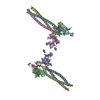 7wz8MC M: atomic model generated by this map C: citing same article ( |
|---|---|
| Similar structure data |
-Links
| EMDB pages | EMDB (EBI/PDBe) / EMDataResource |
|---|---|
| Related items in Molecule of the Month |


-Map
| File | Download / File: emd_32906.map.gz / Format: CCP4 / Size: 8 MB / Type: IMAGE STORED AS FLOATING POINT NUMBER (4 BYTES) | ||||||||||||||||||||
|---|---|---|---|---|---|---|---|---|---|---|---|---|---|---|---|---|---|---|---|---|---|
| Annotation | Langerin trimer in the Birbeck granule. | ||||||||||||||||||||
| Voxel size | X=Y=Z: 2.67 Å | ||||||||||||||||||||
| Density |
| ||||||||||||||||||||
| Symmetry | Space group: 1 | ||||||||||||||||||||
| Details | EMDB XML:
|
-Supplemental data
-Additional map: Langerin trimer in the Birbeck granule. The second...
| File | emd_32906_additional_1.map | ||||||||||||
|---|---|---|---|---|---|---|---|---|---|---|---|---|---|
| Annotation | Langerin trimer in the Birbeck granule. The second body of multibody refinement. | ||||||||||||
| Projections & Slices |
| ||||||||||||
| Density Histograms |
 Z
Z Y
Y X
X


-Half map: Half map 2
| File | emd_32906_half_map_1.map | ||||||||||||
|---|---|---|---|---|---|---|---|---|---|---|---|---|---|
| Annotation | Half map 2 | ||||||||||||
| Projections & Slices |
| ||||||||||||
| Density Histograms |


-Half map: Half map 1
| File | emd_32906_half_map_2.map | ||||||||||||
|---|---|---|---|---|---|---|---|---|---|---|---|---|---|
| Annotation | Half map 1 | ||||||||||||
| Projections & Slices |
| ||||||||||||
| Density Histograms |


- Sample components
-Entire : Repeating unit of langerin lattice in Birbeck granule
| Entire | Name: Repeating unit of langerin lattice in Birbeck granule |
|---|---|
| Components |
|
-Supramolecule #1: Repeating unit of langerin lattice in Birbeck granule
| Supramolecule | Name: Repeating unit of langerin lattice in Birbeck granule / type: complex / ID: 1 / Parent: 0 / Macromolecule list: all Details: Birbeck granules isolated from 293T cells by immunoprecipitation. |
|---|---|
| Source (natural) | Organism: Homo sapiens (human) |
-Macromolecule #1: SNAP-tag,C-type lectin domain family 4 member K
| Macromolecule | Name: SNAP-tag,C-type lectin domain family 4 member K / type: protein_or_peptide / ID: 1 Details: 185 amino acids SNAP-tag + 3xHA tag (YPYDVPDYAYPYDVPDYAYPYDVPDYA) + Linker (GSSG) + HRV3C cleavage site (LEVLFQGP) Number of copies: 6 / Enantiomer: LEVO |
|---|---|
| Source (natural) | Organism: Homo sapiens (human) |
| Molecular weight | Theoretical: 60.547773 KDa |
| Recombinant expression | Organism: Homo sapiens (human) |
| Sequence | String: MDKDCEMKRT TLDSPLGKLE LSGCEQGLHE IKLLGKGTSA ADAVEVPAPA AVLGGPEPLM QATAWLNAYF HQPEAIEEFP VPALHHPVF QQESFTRQVL WKLLKVVKFG EVISYQQLAA LAGNPAATAA VKTALSGNPV PILIPCHRVV SSSGAVGGYE G GLAVKEWL ...String: MDKDCEMKRT TLDSPLGKLE LSGCEQGLHE IKLLGKGTSA ADAVEVPAPA AVLGGPEPLM QATAWLNAYF HQPEAIEEFP VPALHHPVF QQESFTRQVL WKLLKVVKFG EVISYQQLAA LAGNPAATAA VKTALSGNPV PILIPCHRVV SSSGAVGGYE G GLAVKEWL LAHEGHRLGK PGLGPAGYPY DVPDYAYPYD VPDYAYPYDV PDYAGSSGLE VLFQGPTVEK EAPDAHFTVD KQ NISLWPR EPPPKSGPSL VPGKTPTVRA ALICLTLVLV ASVLLQAVLY PRFMGTISDV KTNVQLLKGR VDNISTLDSE IKK NSDGME AAGVQIQMVN ESLGYVRSQF LKLKTSVEKA NAQIQILTRS WEEVSTLNAQ IPELKSDLEK ASALNTKIRA LQGS LENMS KLLKRQNDIL QVVSQGWKYF KGNFYYFSLI PKTWYSAEQF CVSRNSHLTS VTSESEQEFL YKTAGGLIYW IGLTK AGME GDWSWVDDTP FNKVQSARFW IPGEPNNAGN NEHCGNIKAP SLQAWNDAPC DKTFLFICKR PYVPSEP UniProtKB: C-type lectin domain family 4 member K |
-Experimental details
-Structure determination
| Method | cryo EM |
|---|---|
 Processing Processing | subtomogram averaging |
| Aggregation state | 2D array |
-Sample preparation
| Concentration | 0.05 mg/mL |
|---|---|
| Buffer | pH: 7.2 |
| Grid | Model: Quantifoil R1.2/1.3 / Material: COPPER/RHODIUM / Mesh: 200 / Support film - Material: CARBON / Support film - topology: HOLEY / Pretreatment - Type: GLOW DISCHARGE / Pretreatment - Time: 60 sec. / Pretreatment - Atmosphere: AIR |
| Vitrification | Cryogen name: ETHANE / Chamber humidity: 100 % / Chamber temperature: 277 K / Instrument: FEI VITROBOT MARK IV / Details: Blot for 10 seconds before plunging. |
- Electron microscopy
| Microscope | TFS KRIOS |
|---|---|
| Electron beam | Acceleration voltage: 300 kV / Electron source: FIELD EMISSION GUN |
| Electron optics | Calibrated defocus max: 8.5 µm / Calibrated defocus min: 2.6 µm / Illumination mode: FLOOD BEAM / Imaging mode: BRIGHT FIELDBright-field microscopy / Cs: 2.7 mm / Nominal defocus max: 6.0 µm / Nominal defocus min: 3.0 µm / Nominal magnification: 35000 |
| Specialist optics | Energy filter - Name: GIF Quantum LS / Energy filter - Slit width: 20 eV |
| Sample stage | Specimen holder model: FEI TITAN KRIOS AUTOGRID HOLDER / Cooling holder cryogen: NITROGEN |
| Image recording | Film or detector model: GATAN K3 (6k x 4k) / Number grids imaged: 1 / Average exposure time: 0.74 sec. / Average electron dose: 1.24 e/Å2 |
| Experimental equipment |  Model: Titan Krios / Image courtesy: FEI Company |
-Image processing
| Extraction | Number tomograms: 33 / Number images used: 93953 / Software: (Name: IMOD (ver. 4.11.11), RELION (ver. 4.0)) |
|---|---|
| Final 3D classification | Software - Name: RELION (ver. 4.0) |
| Final angle assignment | Type: ANGULAR RECONSTITUTION / Software - Name: RELION (ver. 4.0) |
| Final reconstruction | Number classes used: 1 / Applied symmetry - Point group: C3 (3 fold cyclic) / Resolution.type: BY AUTHOR / Resolution: 6.4 Å / Resolution method: FSC 0.143 CUT-OFF / Software - Name: RELION (ver. 4.0) / Number subtomograms used: 63563 |
| FSC plot (resolution estimation) |  |

