Movie
Movie Controller

[English] 日本語
 Yorodumi
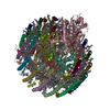
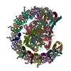
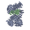
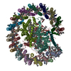
- EMDB-31400: STRUCTURE OF PHOTOSYNTHETIC LH1-RC SUPER-COMPLEX OF RHODOBACTER S... -
+ Open data

- Basic information
| Entry | Database: EMDB / ID: EMD-31400 | |||||||||||||||||||||
|---|---|---|---|---|---|---|---|---|---|---|---|---|---|---|---|---|---|---|---|---|---|---|
| Title | STRUCTURE OF PHOTOSYNTHETIC LH1-RC SUPER-COMPLEX OF RHODOBACTER SPHAEROIDES MONOMER | |||||||||||||||||||||
 Map data Map data | ||||||||||||||||||||||
 Sample Sample |
| |||||||||||||||||||||
| Function / homology |  Function and homology information Function and homology informationorganelle inner membrane / plasma membrane light-harvesting complex /  bacteriochlorophyll binding / photosynthesis, light reaction / electron transporter, transferring electrons within the cyclic electron transport pathway of photosynthesis activity / membrane => GO:0016020 / metal ion binding / plasma membrane bacteriochlorophyll binding / photosynthesis, light reaction / electron transporter, transferring electrons within the cyclic electron transport pathway of photosynthesis activity / membrane => GO:0016020 / metal ion binding / plasma membraneSimilarity search - Function | |||||||||||||||||||||
| Biological species |  Rhodobacter sphaeroides f. sp. denitrificans (bacteria) / Rhodobacter sphaeroides (bacteria) / Rhodopseudomonas sphaeroides (bacteria) Rhodobacter sphaeroides f. sp. denitrificans (bacteria) / Rhodobacter sphaeroides (bacteria) / Rhodopseudomonas sphaeroides (bacteria) | |||||||||||||||||||||
| Method | single particle reconstruction / cryo EM / Resolution: 2.94 Å | |||||||||||||||||||||
 Authors Authors | Tani K / Nagashima VP / Kanno R / Kawamura S / Kikuchi R / Ji X-C / Hall M / Yu L-J / Kimura Y / Madigan MT ...Tani K / Nagashima VP / Kanno R / Kawamura S / Kikuchi R / Ji X-C / Hall M / Yu L-J / Kimura Y / Madigan MT / Mizoguchi A / Humbel BM / Wang-Otomo Z-Y | |||||||||||||||||||||
| Funding support |  Japan, 6 items Japan, 6 items
| |||||||||||||||||||||
 Citation Citation | Journal: Nat Commun / Year: 2021 Title: A previously unrecognized membrane protein in the Rhodobacter sphaeroides LH1-RC photocomplex. Authors: Kazutoshi Tani / Kenji V P Nagashima / Ryo Kanno / Saki Kawamura / Riku Kikuchi / Malgorzata Hall / Long-Jiang Yu / Yukihiro Kimura / Michael T Madigan / Akira Mizoguchi / Bruno M Humbel / ...Authors: Kazutoshi Tani / Kenji V P Nagashima / Ryo Kanno / Saki Kawamura / Riku Kikuchi / Malgorzata Hall / Long-Jiang Yu / Yukihiro Kimura / Michael T Madigan / Akira Mizoguchi / Bruno M Humbel / Zheng-Yu Wang-Otomo /   Abstract: Rhodobacter (Rba.) sphaeroides is the most widely used model organism in bacterial photosynthesis. The light-harvesting-reaction center (LH1-RC) core complex of this purple phototroph is ...Rhodobacter (Rba.) sphaeroides is the most widely used model organism in bacterial photosynthesis. The light-harvesting-reaction center (LH1-RC) core complex of this purple phototroph is characterized by the co-existence of monomeric and dimeric forms, the presence of the protein PufX, and approximately two carotenoids per LH1 αβ-polypeptides. Despite many efforts, structures of the Rba. sphaeroides LH1-RC have not been obtained at high resolutions. Here we report a cryo-EM structure of the monomeric LH1-RC from Rba. sphaeroides strain IL106 at 2.9 Å resolution. The LH1 complex forms a C-shaped structure composed of 14 αβ-polypeptides around the RC with a large ring opening. From the cryo-EM density map, a previously unrecognized integral membrane protein, referred to as protein-U, was identified. Protein-U has a U-shaped conformation near the LH1-ring opening and was annotated as a hypothetical protein in the Rba. sphaeroides genome. Deletion of protein-U resulted in a mutant strain that expressed a much-reduced amount of the dimeric LH1-RC, indicating an important role for protein-U in dimerization of the LH1-RC complex. PufX was located opposite protein-U on the LH1-ring opening, and both its position and conformation differed from that of previous reports of dimeric LH1-RC structures obtained at low-resolution. Twenty-six molecules of the carotenoid spheroidene arranged in two distinct configurations were resolved in the Rba. sphaeroides LH1 and were positioned within the complex to block its channels. Our findings offer an exciting new view of the core photocomplex of Rba. sphaeroides and the connections between structure and function in bacterial photocomplexes in general. | |||||||||||||||||||||
| History |
|
- Structure visualization
| Movie |
Movie viewer |
|---|---|
| Structure viewer | EM map: SurfViewMolmilJmol/JSmol |
| Supplemental images |

- Downloads & links
-EMDB archive
| Map data | emd_31400.map.gz | 78.3 MB | EMDB map data format | |
|---|---|---|---|---|
| Header (meta data) | emd-31400-v30.xmlemd-31400.xml | 23.1 KB 23.1 KB | Display Display | EMDB header |
| FSC (resolution estimation) | emd_31400_fsc.xml | 9.9 KB | Display | FSC data file |
| Images | 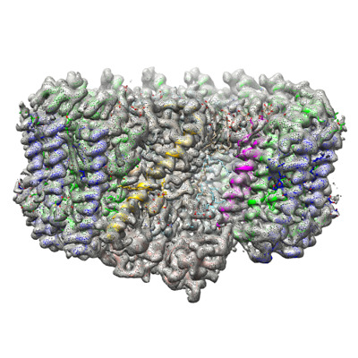 emd_31400.png emd_31400.png | 187.2 KB | ||
| Archive directory |  http://ftp.pdbj.org/pub/emdb/structures/EMD-31400ftp://ftp.pdbj.org/pub/emdb/structures/EMD-31400 http://ftp.pdbj.org/pub/emdb/structures/EMD-31400ftp://ftp.pdbj.org/pub/emdb/structures/EMD-31400 | HTTPS FTP |
-Related structure data
| Related structure data | 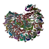 7f0lMC M: atomic model generated by this map C: citing same article ( |
|---|---|
| Similar structure data |


-Links
| EMDB pages | EMDB (EBI/PDBe) / EMDataResource |
|---|
-Map
| File | Download / File: emd_31400.map.gz / Format: CCP4 / Size: 83.7 MB / Type: IMAGE STORED AS FLOATING POINT NUMBER (4 BYTES) | ||||||||||||||||||||||||||||||||||||||||||||||||||||||||||||||||||||
|---|---|---|---|---|---|---|---|---|---|---|---|---|---|---|---|---|---|---|---|---|---|---|---|---|---|---|---|---|---|---|---|---|---|---|---|---|---|---|---|---|---|---|---|---|---|---|---|---|---|---|---|---|---|---|---|---|---|---|---|---|---|---|---|---|---|---|---|---|---|
| Voxel size | X=Y=Z: 1.094 Å | ||||||||||||||||||||||||||||||||||||||||||||||||||||||||||||||||||||
| Density |
| ||||||||||||||||||||||||||||||||||||||||||||||||||||||||||||||||||||
| Symmetry | Space group: 1 | ||||||||||||||||||||||||||||||||||||||||||||||||||||||||||||||||||||
| Details | EMDB XML:
CCP4 map header:
| ||||||||||||||||||||||||||||||||||||||||||||||||||||||||||||||||||||
-Supplemental data
- Sample components
+Entire : Photosynthetic LH1-RC complex from the purple phototrophic bacter...
+Supramolecule #1: Photosynthetic LH1-RC complex from the purple phototrophic bacter...
+Macromolecule #1: Photosynthetic reaction center L subunit
+Macromolecule #2: Reaction center protein M chain
+Macromolecule #3: Reaction center protein H chain
+Macromolecule #4: Light-harvesting protein B-875 alpha chain
+Macromolecule #5: Antenna pigment protein beta chain
+Macromolecule #6: Light-harvesting protein B-875 alpha chain
+Macromolecule #7: PufX
+Macromolecule #8: protein-U
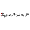
+Macromolecule #9: BACTERIOCHLOROPHYLL A

+Macromolecule #10: BACTERIOPHEOPHYTIN A

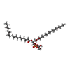
+Macromolecule #11: UBIQUINONE-10
+Macromolecule #12: (1R)-2-{[{[(2S)-2,3-DIHYDROXYPROPYL]OXY}(HYDROXY)PHOSPHORYL]OXY}-...
+Macromolecule #13: LAURYL DIMETHYLAMINE-N-OXIDE

+Macromolecule #14: FE (III) ION
+Macromolecule #15: SPHEROIDENE
+Macromolecule #16: DODECYL-BETA-D-MALTOSIDE

+Macromolecule #17: CARDIOLIPIN
-Experimental details
-Structure determination
| Method | cryo EM |
|---|---|
 Processing Processing | single particle reconstruction |
| Aggregation state | particle |
-Sample preparation
| Concentration | 3.0 mg/mL |
|---|---|
| Buffer | pH: 8 |
| Grid | Material: MOLYBDENUM |
| Vitrification | Cryogen name: ETHANE / Chamber humidity: 80 % / Chamber temperature: 277 K / Instrument: LEICA EM GP |
| Details | This sample was monodisperse. |
- Electron microscopy
| Microscope | FEI TALOS ARCTICA |
|---|---|
| Electron beam | Acceleration voltage: 200 kV / Electron source: FIELD EMISSION GUN |
| Electron optics | Illumination mode: FLOOD BEAM / Imaging mode: BRIGHT FIELDBright-field microscopy |
| Sample stage | Specimen holder model: FEI TITAN KRIOS AUTOGRID HOLDER / Cooling holder cryogen: NITROGEN |
| Image recording | Film or detector model: FEI FALCON III (4k x 4k) / Detector mode: COUNTING / Average exposure time: 1.275 sec. / Average electron dose: 42.0 e/Å2 |
| Experimental equipment | 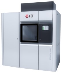 Model: Talos Arctica / Image courtesy: FEI Company |
-Image processing
| Particle selection | Number selected: 551846 |
|---|---|
| CTF correction | Software - Name: CTFFIND |
| Initial angle assignment | Type: NOT APPLICABLE |
| Final 3D classification | Number classes: 4 / Avg.num./class: 160448 / Software - Name: RELION (ver. 3.1) |
| Final angle assignment | Type: MAXIMUM LIKELIHOOD / Software - Name: RELION (ver. 3.1) |
| Final reconstruction | Resolution.type: BY AUTHOR / Resolution: 2.94 Å / Resolution method: FSC 0.143 CUT-OFF / Software - Name: RELION (ver. 3.1) / Number images used: 160448 |
| FSC plot (resolution estimation) |  |