 Movie
Movie Controller

[English] 日本語
 Yorodumi
- EMDB-30306: Structural basis for cross-species recognition of COVID-19 virus ... -
+ Open data

- Basic information
| Entry | Database: EMDB / ID: EMD-30306 | |||||||||
|---|---|---|---|---|---|---|---|---|---|---|
| Title | Structural basis for cross-species recognition of COVID-19 virus spike receptor binding domain to bat ACE2 | |||||||||
 Map data Map data | Cryo-EM structure of bat ACE2 | |||||||||
 Sample Sample |
| |||||||||
| Function / homology |  Function and homology information Function and homology information Hydrolases; Acting on peptide bonds (peptidases) / peptidyl-dipeptidase activity / carboxypeptidase activity / cilium / metallopeptidase activity / apical plasma membrane / proteolysis / extracellular region / metal ion binding / cytoplasm Hydrolases; Acting on peptide bonds (peptidases) / peptidyl-dipeptidase activity / carboxypeptidase activity / cilium / metallopeptidase activity / apical plasma membrane / proteolysis / extracellular region / metal ion binding / cytoplasmSimilarity search - Function | |||||||||
| Biological species |  Bat AAV SC2991 (virus) / Bat AAV SC2991 (virus) /  Rhinolophus macrotis (Big-eared Horseshoe Bat) Rhinolophus macrotis (Big-eared Horseshoe Bat) | |||||||||
| Method | single particle reconstruction / cryo EM / Resolution: 3.2 Å | |||||||||
 Authors Authors | Liu KF / Wang J / Tan SG / Niu S / Wu LL / Zhang YF / Pan XQ / Meng YM / Chen Q / Wang QH ...Liu KF / Wang J / Tan SG / Niu S / Wu LL / Zhang YF / Pan XQ / Meng YM / Chen Q / Wang QH / Wang HW / Qi JX / Gao GF | |||||||||
 Citation Citation | Journal: Proc Natl Acad Sci U S A / Year: 2021 Title: Cross-species recognition of SARS-CoV-2 to bat ACE2. Authors: Kefang Liu / Shuguang Tan / Sheng Niu / Jia Wang / Lili Wu / Huan Sun / Yanfang Zhang / Xiaoqian Pan / Xiao Qu / Pei Du / Yumin Meng / Yunfei Jia / Qian Chen / Chuxia Deng / Jinghua Yan / ...Authors: Kefang Liu / Shuguang Tan / Sheng Niu / Jia Wang / Lili Wu / Huan Sun / Yanfang Zhang / Xiaoqian Pan / Xiao Qu / Pei Du / Yumin Meng / Yunfei Jia / Qian Chen / Chuxia Deng / Jinghua Yan / Hong-Wei Wang / Qihui Wang / Jianxun Qi / George Fu Gao /  Abstract: The coronavirus disease 2019 (COVID-19) pandemic caused by severe acute respiratory syndrome coronavirus 2 (SARS-CoV-2) has emerged as a major threat to global health. Although varied SARS-CoV-2- ...The coronavirus disease 2019 (COVID-19) pandemic caused by severe acute respiratory syndrome coronavirus 2 (SARS-CoV-2) has emerged as a major threat to global health. Although varied SARS-CoV-2-related coronaviruses have been isolated from bats and SARS-CoV-2 may infect bat, the structural basis for SARS-CoV-2 to utilize the human receptor counterpart bat angiotensin-converting enzyme 2 (bACE2) for virus infection remains less understood. Here, we report that the SARS-CoV-2 spike protein receptor binding domain (RBD) could bind to bACE2 from (bACE2-Rm) with substantially lower affinity compared with that to the human ACE2 (hACE2), and its infectivity to host cells expressing bACE2-Rm was confirmed with pseudotyped SARS-CoV-2 virus and SARS-CoV-2 wild virus. The structure of the SARS-CoV-2 RBD with the bACE2-Rm complex was determined, revealing a binding mode similar to that of hACE2. The analysis of binding details between SARS-CoV-2 RBD and bACE2-Rm revealed that the interacting network involving Y41 and E42 of bACE2-Rm showed substantial differences with that to hACE2. Bats have extensive species diversity and the residues for RBD binding in bACE2 receptor varied substantially among different bat species. Notably, the Y41H mutant, which exists in many bats, attenuates the binding capacity of bACE2-Rm, indicating the central roles of Y41 in the interaction network. These findings would benefit our understanding of the potential infection of SARS-CoV-2 in varied species of bats. | |||||||||
| History |
|
- Structure visualization
| Movie |
Movie viewer |
|---|---|
| Structure viewer | EM map: SurfViewMolmilJmol/JSmol |
| Supplemental images |

- Downloads & links
-EMDB archive
| Map data | emd_30306.map.gz | 1.5 MB | EMDB map data format | |
|---|---|---|---|---|
| Header (meta data) | emd-30306-v30.xmlemd-30306.xml | 10.9 KB 10.9 KB | Display Display | EMDB header |
| FSC (resolution estimation) | emd_30306_fsc.xml | 5.8 KB | Display | FSC data file |
| Images | 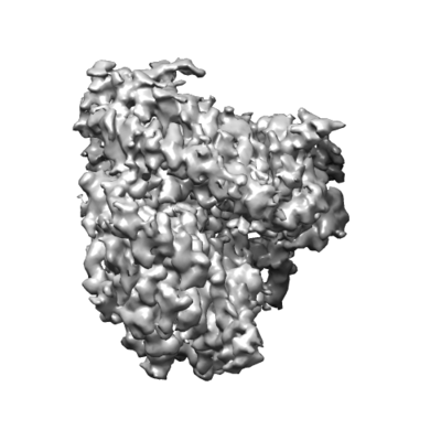 emd_30306.png emd_30306.png | 56.3 KB | ||
| Archive directory |  http://ftp.pdbj.org/pub/emdb/structures/EMD-30306ftp://ftp.pdbj.org/pub/emdb/structures/EMD-30306 http://ftp.pdbj.org/pub/emdb/structures/EMD-30306ftp://ftp.pdbj.org/pub/emdb/structures/EMD-30306 | HTTPS FTP |
-Related structure data
| Related structure data |  7c8kMC  7c8jC M: atomic model generated by this map C: citing same article ( |
|---|---|
| Similar structure data |


-Links
| EMDB pages | EMDB (EBI/PDBe) / EMDataResource |
|---|---|
| Related items in Molecule of the Month |


-Map
| File | Download / File: emd_30306.map.gz / Format: CCP4 / Size: 15.6 MB / Type: IMAGE STORED AS FLOATING POINT NUMBER (4 BYTES) | ||||||||||||||||||||||||||||||||||||||||||||||||||||||||||||||||||||
|---|---|---|---|---|---|---|---|---|---|---|---|---|---|---|---|---|---|---|---|---|---|---|---|---|---|---|---|---|---|---|---|---|---|---|---|---|---|---|---|---|---|---|---|---|---|---|---|---|---|---|---|---|---|---|---|---|---|---|---|---|---|---|---|---|---|---|---|---|---|
| Annotation | Cryo-EM structure of bat ACE2 | ||||||||||||||||||||||||||||||||||||||||||||||||||||||||||||||||||||
| Voxel size | X=Y=Z: 0.99375 Å | ||||||||||||||||||||||||||||||||||||||||||||||||||||||||||||||||||||
| Density |
| ||||||||||||||||||||||||||||||||||||||||||||||||||||||||||||||||||||
| Symmetry | Space group: 1 | ||||||||||||||||||||||||||||||||||||||||||||||||||||||||||||||||||||
| Details | EMDB XML:
CCP4 map header:
| ||||||||||||||||||||||||||||||||||||||||||||||||||||||||||||||||||||
-Supplemental data
- Sample components
-Entire : Bat ACE2
| Entire | Name: Bat ACE2 |
|---|---|
| Components |
|
-Supramolecule #1: Bat ACE2
| Supramolecule | Name: Bat ACE2 / type: cell / ID: 1 / Parent: 0 / Macromolecule list: #1 |
|---|---|
| Source (natural) | Organism: Bat AAV SC2991 (virus) |
-Macromolecule #1: Angiotensin-converting enzyme
| Macromolecule | Name: Angiotensin-converting enzyme / type: protein_or_peptide / ID: 1 / Number of copies: 1 / Enantiomer: LEVO / EC number: Hydrolases; Acting on peptide bonds (peptidases) |
|---|---|
| Source (natural) | Organism: Rhinolophus macrotis (Big-eared Horseshoe Bat) |
| Molecular weight | Theoretical: 69.444312 KDa |
| Recombinant expression | Organism:  Homo sapiens (human) Homo sapiens (human) |
| Sequence | String: STTEDEAKKF LDKFNSKAED LSYESSLASW DYNTNISDEN VQKMDEAGAK WSAFYEEQSK LAKNYPLEEI QNDTVKRQLQ ILQQSGSPV LSEDKSKRLN SILNAMSTIY STGKVCKPNN PQECLLLEPG LDNIMGTSKD YNERLWAWEG WRAEVGKQLR P LYEEYVVL ...String: STTEDEAKKF LDKFNSKAED LSYESSLASW DYNTNISDEN VQKMDEAGAK WSAFYEEQSK LAKNYPLEEI QNDTVKRQLQ ILQQSGSPV LSEDKSKRLN SILNAMSTIY STGKVCKPNN PQECLLLEPG LDNIMGTSKD YNERLWAWEG WRAEVGKQLR P LYEEYVVL KNEMARGYHY EDYGDYWRRD YETEESSGPG YSRDQLMKDV DRIFTEIKPL YEHLHAYVRA KLMDTYPLHI SP TGCLPAH LLGDMWGRFW TNLYPLTVPF GQKPNIDVTD AMLNQGWDAN RIFKEAEKFF VSVSLPKMTE GFWNKSMLTE PGD GRKVVC HPTAWDLGKG DFRIKMCTKV TMEDFLTAHH EMGHIQYDMA YASQPYLLRN GANEGFHEAV GEVMSLSVAT PKHL KTMGL LSPDFREDDE TEINFLLKQA LNIVGTLPFT YMLEKWRWMV FKGEIPKEEW MKKWWEMKRE IVGVVEPVPH DETYC DPAS LFHVANDYSF IRYYTRTIFE FQFHEALCRI AQHNGPLHKC DISNSTDAGK KLHQMLSVGK SQAWTKTLED IVDSRN MDV GPLLRYFKPL YTWLQEQNRK SYVGWNTDWS PYA |
-Macromolecule #3: 2-acetamido-2-deoxy-beta-D-glucopyranose
| Macromolecule | Name: 2-acetamido-2-deoxy-beta-D-glucopyranose / type: ligand / ID: 3 / Number of copies: 3 / Formula: NAG |
|---|---|
| Molecular weight | Theoretical: 221.208 Da |
| Chemical component information |  ChemComp-NAG: |
-Macromolecule #4: ZINC ION
| Macromolecule | Name: ZINC ION / type: ligand / ID: 4 / Number of copies: 1 / Formula: ZN |
|---|---|
| Molecular weight | Theoretical: 65.409 Da |
-Experimental details
-Structure determination
| Method | cryo EM |
|---|---|
 Processing Processing | single particle reconstruction |
| Aggregation state | particle |
-Sample preparation
| Buffer | pH: 8 |
|---|---|
| Vitrification | Cryogen name: ETHANE |
- Electron microscopy
| Microscope | FEI TITAN KRIOS |
|---|---|
| Electron beam | Acceleration voltage: 300 kV / Electron source: FIELD EMISSION GUN |
| Electron optics | Illumination mode: FLOOD BEAM / Imaging mode: BRIGHT FIELDBright-field microscopy |
| Image recording | Film or detector model: GATAN K3 BIOQUANTUM (6k x 4k) / Average electron dose: 50.0 e/Å2 |
| Experimental equipment |  Model: Titan Krios / Image courtesy: FEI Company |
-Image processing
| Initial angle assignment | Type: MAXIMUM LIKELIHOOD |
|---|---|
| Final angle assignment | Type: MAXIMUM LIKELIHOOD |
| Final reconstruction | Applied symmetry - Point group: C1 (asymmetric) / Resolution.type: BY AUTHOR / Resolution: 3.2 Å / Resolution method: FSC 0.143 CUT-OFF / Number images used: 62289 |
| FSC plot (resolution estimation) |  |
