[English] 日本語
Yorodumi
- EMDB-29016: Cryo-EM structure of SARS-CoV-2 postfusion spike in membrane -

+
Open data


ID or keywords:

Loading...

-
Basic information

Entry
Database: EMDB / ID: EMD-29016
TitleCryo-EM structure of SARS-CoV-2 postfusion spike in membrane
Map data
Sample
  • Complex: SRAS-CoV-2 postfusion spike protein in nanodisc
    • Protein or peptide: Spike protein S2
  • Ligand: alpha-D-mannopyranose
  • Ligand: 2-acetamido-2-deoxy-beta-D-glucopyranose
KeywordsVIRAL PROTEIN
Function / homology
Function and homology information


Maturation of spike protein / viral translation / Translation of Structural Proteins / Virion Assembly and Release / host cell surface / host extracellular space / suppression by virus of host tetherin activity / Induction of Cell-Cell Fusion / structural constituent of virion / entry receptor-mediated virion attachment to host cell ...Maturation of spike protein / viral translation / Translation of Structural Proteins / Virion Assembly and Release / host cell surface / host extracellular space / suppression by virus of host tetherin activity / Induction of Cell-Cell Fusion / structural constituent of virion / entry receptor-mediated virion attachment to host cell / host cell endoplasmic reticulum-Golgi intermediate compartment membrane / receptor-mediated endocytosis of virus by host cell / Attachment and Entry / membrane fusion / positive regulation of viral entry into host cell / receptor-mediated virion attachment to host cell / receptor ligand activity / host cell surface receptor binding / fusion of virus membrane with host plasma membrane / fusion of virus membrane with host endosome membrane / viral envelope / virion attachment to host cell / SARS-CoV-2 activates/modulates innate and adaptive immune responses / host cell plasma membrane / virion membrane / membrane / identical protein binding / plasma membrane
Similarity search - Function
Spike (S) protein S1 subunit, receptor-binding domain, SARS-CoV-2 / Spike (S) protein S1 subunit, N-terminal domain, SARS-CoV-like / Betacoronavirus spike (S) glycoprotein S1 subunit N-terminal (NTD) domain profile. / Spike glycoprotein, N-terminal domain superfamily / Betacoronavirus spike (S) glycoprotein S1 subunit C-terminal (CTD) domain profile. / Spike glycoprotein, betacoronavirus / Spike (S) protein S1 subunit, receptor-binding domain, betacoronavirus / Spike S1 subunit, receptor binding domain superfamily, betacoronavirus / Betacoronavirus spike glycoprotein S1, receptor binding / Spike glycoprotein S1, N-terminal domain, betacoronavirus-like ...Spike (S) protein S1 subunit, receptor-binding domain, SARS-CoV-2 / Spike (S) protein S1 subunit, N-terminal domain, SARS-CoV-like / Betacoronavirus spike (S) glycoprotein S1 subunit N-terminal (NTD) domain profile. / Spike glycoprotein, N-terminal domain superfamily / Betacoronavirus spike (S) glycoprotein S1 subunit C-terminal (CTD) domain profile. / Spike glycoprotein, betacoronavirus / Spike (S) protein S1 subunit, receptor-binding domain, betacoronavirus / Spike S1 subunit, receptor binding domain superfamily, betacoronavirus / Betacoronavirus spike glycoprotein S1, receptor binding / Spike glycoprotein S1, N-terminal domain, betacoronavirus-like / Betacoronavirus-like spike glycoprotein S1, N-terminal / Spike glycoprotein S2, coronavirus, heptad repeat 1 / Spike glycoprotein S2, coronavirus, heptad repeat 2 / Coronavirus spike (S) glycoprotein S2 subunit heptad repeat 2 (HR2) region profile. / Coronavirus spike (S) glycoprotein S2 subunit heptad repeat 1 (HR1) region profile. / Spike glycoprotein S2 superfamily, coronavirus / Spike glycoprotein S2, coronavirus / Coronavirus spike glycoprotein S2 / Coronavirus spike glycoprotein S1, C-terminal / Coronavirus spike glycoprotein S1, C-terminal
Similarity search - Domain/homology
Biological speciesSevere acute respiratory syndrome coronavirus 2 / Severe acute respiratory syndrome coronavirus
Methodsingle particle reconstruction / cryo EM / Resolution: 2.9 Å
AuthorsZhang J / Shi W / Cai YF / Zhu HS / Peng HQ / Voyer J / Volloch SR / Cao H / Mayer ML / Song KK ...Zhang J / Shi W / Cai YF / Zhu HS / Peng HQ / Voyer J / Volloch SR / Cao H / Mayer ML / Song KK / Xu C / Lu JM / Chen B
Funding support United States, 3 items
OrganizationGrant numberCountry
National Institutes of Health/National Institute Of Allergy and Infectious Diseases (NIH/NIAID)AI147884 United States
National Institutes of Health/National Institute Of Allergy and Infectious Diseases (NIH/NIAID)AI141002 United States
National Institutes of Health/National Institute Of Allergy and Infectious Diseases (NIH/NIAID)AI127193 United States
Citation
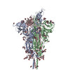
Journal: Nature / Year: 2023
Title: Cryo-EM structure of SARS-CoV-2 postfusion spike in membrane.
Authors: Wei Shi / Yongfei Cai / Haisun Zhu / Hanqin Peng / Jewel Voyer / Sophia Rits-Volloch / Hong Cao / Megan L Mayer / Kangkang Song / Chen Xu / Jianming Lu / Jun Zhang / Bing Chen /
Abstract: The entry of SARS-CoV-2 into host cells depends on the refolding of the virus-encoded spike protein from a prefusion conformation, which is metastable after cleavage, to a lower-energy stable ...The entry of SARS-CoV-2 into host cells depends on the refolding of the virus-encoded spike protein from a prefusion conformation, which is metastable after cleavage, to a lower-energy stable postfusion conformation. This transition overcomes kinetic barriers for fusion of viral and target cell membranes. Here we report a cryogenic electron microscopy (cryo-EM) structure of the intact postfusion spike in a lipid bilayer that represents the single-membrane product of the fusion reaction. The structure provides structural definition of the functionally critical membrane-interacting segments, including the fusion peptide and transmembrane anchor. The internal fusion peptide forms a hairpin-like wedge that spans almost the entire lipid bilayer and the transmembrane segment wraps around the fusion peptide at the last stage of membrane fusion. These results advance our understanding of the spike protein in a membrane environment and may guide development of intervention strategies.
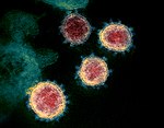
History
DepositionDec 5, 2022-
Header (metadata) releaseMay 10, 2023-
Map releaseMay 10, 2023-
UpdateJul 26, 2023-
Current statusJul 26, 2023Processing site: RCSB / Status: Released

-
Structure visualization

Supplemental images

Downloads & links

-
Map

FileDownload / File: emd_29016.map.gz / Format: CCP4 / Size: 824 MB / Type: IMAGE STORED AS FLOATING POINT NUMBER (4 BYTES)
Voxel sizeX=Y=Z: 0.825 Å
Density
Contour LevelBy AUTHOR: 0.15
Minimum - Maximum-0.002663985 - 2.532832
Average (Standard dev.)0.0011990807 (±0.02302369)
SymmetrySpace group: 1
Details

EMDB XML:

Map geometry
Axis orderXYZ
Origin000
Dimensions600600600
Spacing600600600
CellA=B=C: 495.0 Å
α=β=γ: 90.0 °

-
Supplemental data

-
Half map: #2

Fileemd_29016_half_map_1.map
Projections & Slices
AxesZYX

Projections

Slices (1/2)
Density Histograms

-
Half map: #1

Fileemd_29016_half_map_2.map
Projections & Slices
AxesZYX

Projections

Slices (1/2)
Density Histograms

-
Sample components

-
Entire : SRAS-CoV-2 postfusion spike protein in nanodisc

EntireName: SRAS-CoV-2 postfusion spike protein in nanodisc
Components
  • Complex: SRAS-CoV-2 postfusion spike protein in nanodisc
    • Protein or peptide: Spike protein S2
  • Ligand: alpha-D-mannopyranose
  • Ligand: 2-acetamido-2-deoxy-beta-D-glucopyranose

-
Supramolecule #1: SRAS-CoV-2 postfusion spike protein in nanodisc

SupramoleculeName: SRAS-CoV-2 postfusion spike protein in nanodisc / type: complex / ID: 1 / Parent: 0 / Macromolecule list: #1 / Details: SARS-CoV-2 postfusion spike protein in nanodisc
Source (natural)Organism: Severe acute respiratory syndrome coronavirus 2
Molecular weightTheoretical: 210 KDa

-
Macromolecule #1: Spike protein S2

MacromoleculeName: Spike protein S2 / type: protein_or_peptide / ID: 1 / Number of copies: 3 / Enantiomer: LEVO
Source (natural)Organism: Severe acute respiratory syndrome coronavirus
Molecular weightTheoretical: 68.176188 KDa
Recombinant expressionOrganism: Homo sapiens (human)
SequenceString: SVASQSIIAY TMSLGAENSV AYSNNSIAIP TNFTISVTTE ILPVSMTKTS VDCTMYICGD STECSNLLLQ YGSFCTQLNR ALTGIAVEQ DKNTQEVFAQ VKQIYKTPPI KDFGGFNFSQ ILPDPSKPSK RSFIEDLLFN KVTLADAGFI KQYGDCLGDI A ARDLICAQ ...String:
SVASQSIIAY TMSLGAENSV AYSNNSIAIP TNFTISVTTE ILPVSMTKTS VDCTMYICGD STECSNLLLQ YGSFCTQLNR ALTGIAVEQ DKNTQEVFAQ VKQIYKTPPI KDFGGFNFSQ ILPDPSKPSK RSFIEDLLFN KVTLADAGFI KQYGDCLGDI A ARDLICAQ KFNGLTVLPP LLTDEMIAQY TSALLAGTIT SGWTFGAGAA LQIPFAMQMA YRFNGIGVTQ NVLYENQKLI AN QFNSAIG KIQDSLSSTA SALGKLQDVV NQNAQALNTL VKQLSSNFGA ISSVLNDILS RLDKVEAEVQ IDRLITGRLQ SLQ TYVTQQ LIRAAEIRAS ANLAATKMSE CVLGQSKRVD FCGKGYHLMS FPQSAPHGVV FLHVTYVPAQ EKNFTTAPAI CHDG KAHFP REGVFVSNGT HWFVTQRNFY EPQIITTDNT FVSGNCDVVI GIVNNTVYDP LQPELDSFKE ELDKYFKNHT SPDVD LGDI SGINASVVNI QKEIDRLNEV AKNLNESLID LQELGKYEQY IKWPWYIWLG FIAGLIAIVM VTIMLCCMTS CCSCLK GCC SCGSCCKFDE DDSEPVLKGV KLHYTLESGG GSAWSHPQFE KGGGSGGGSG GSSAWSHPQF EK

UniProtKB: Spike glycoprotein

-
Macromolecule #5: alpha-D-mannopyranose

MacromoleculeName: alpha-D-mannopyranose / type: ligand / ID: 5 / Number of copies: 3 / Formula: MAN
Molecular weightTheoretical: 180.156 Da
Chemical component information

ChemComp-MAN:
alpha-D-mannopyranose / Mannose

-
Macromolecule #6: 2-acetamido-2-deoxy-beta-D-glucopyranose

MacromoleculeName: 2-acetamido-2-deoxy-beta-D-glucopyranose / type: ligand / ID: 6 / Number of copies: 9 / Formula: NAG
Molecular weightTheoretical: 221.208 Da
Chemical component information

ChemComp-NAG:
2-acetamido-2-deoxy-beta-D-glucopyranose / N-Acetylglucosamine

-
Experimental details

-
Structure determination

Methodcryo EM
Processingsingle particle reconstruction
Aggregation stateparticle

-
Sample preparation

Concentration1.5 mg/mL
BufferpH: 7.5
Component:
ConcentrationFormulaName
0.15 mol/LNaClSodium chloridesodium chloride
0.025 mol/LTris-HClTrisTris hydrochloride
VitrificationCryogen name: ETHANE / Chamber humidity: 100 % / Chamber temperature: 277.15 K / Instrument: FEI VITROBOT MARK IV

-
Electron microscopy

MicroscopeFEI TITAN KRIOS
Electron beamAcceleration voltage: 300 kV / Electron source: FIELD EMISSION GUN
Electron opticsIllumination mode: FLOOD BEAM / Imaging mode: BRIGHT FIELDBright-field microscopy / Nominal defocus max: 2.2 µm / Nominal defocus min: 0.8 µm
Image recordingFilm or detector model: GATAN K3 BIOQUANTUM (6k x 4k) / Average electron dose: 1.01 e/Å2
Experimental equipment
Model: Titan Krios / Image courtesy: FEI Company

-
Image processing

Particle selectionNumber selected: 75996334
Startup modelType of model: PDB ENTRY
PDB model - PDB ID:
Initial angle assignmentType: MAXIMUM LIKELIHOOD
Final 3D classificationSoftware - Name: RELION
Final angle assignmentType: MAXIMUM LIKELIHOOD
Final reconstructionResolution.type: BY AUTHOR / Resolution: 2.9 Å / Resolution method: FSC 0.143 CUT-OFF / Software - Name: cryoSPARC (ver. 4.0.2) / Number images used: 254902

-
Atomic model buiding 1

Initial modelPDB ID:

Chain - Chain ID: A / Chain - Residue range: 14-1211 / Chain - Source name: PDB / Chain - Initial model type: experimental model
RefinementProtocol: AB INITIO MODEL
Output model

PDB-8fdw:
Cryo-EM structure of SARS-CoV-2 postfusion spike in membrane

+
About Yorodumi

-
News

-
Feb 9, 2022. New format data for meta-information of EMDB entries

New format data for meta-information of EMDB entries

  • Version 3 of the EMDB header file is now the official format.
  • The previous official version 1.9 will be removed from the archive.

Related info.:EMDB header

External links:wwPDB to switch to version 3 of the EMDB data model

-
Aug 12, 2020. Covid-19 info

Covid-19 info

URL: https://pdbj.org/emnavi/covid19.php

New page: Covid-19 featured information page in EM Navigator.

Related info.:Covid-19 info / Mar 5, 2020. Novel coronavirus structure data

+
Mar 5, 2020. Novel coronavirus structure data

Novel coronavirus structure data

Related info.:Yorodumi Speices / Aug 12, 2020. Covid-19 info

External links:COVID-19 featured content - PDBj / Molecule of the Month (242):Coronavirus Proteases

+
Jan 31, 2019. EMDB accession codes are about to change! (news from PDBe EMDB page)

EMDB accession codes are about to change! (news from PDBe EMDB page)

  • The allocation of 4 digits for EMDB accession codes will soon come to an end. Whilst these codes will remain in use, new EMDB accession codes will include an additional digit and will expand incrementally as the available range of codes is exhausted. The current 4-digit format prefixed with “EMD-” (i.e. EMD-XXXX) will advance to a 5-digit format (i.e. EMD-XXXXX), and so on. It is currently estimated that the 4-digit codes will be depleted around Spring 2019, at which point the 5-digit format will come into force.
  • The EM Navigator/Yorodumi systems omit the EMD- prefix.

Related info.:Q: What is EMD? / ID/Accession-code notation in Yorodumi/EM Navigator

External links:EMDB Accession Codes are Changing Soon! / Contact to PDBj

+
Jul 12, 2017. Major update of PDB

Major update of PDB

  • wwPDB released updated PDB data conforming to the new PDBx/mmCIF dictionary.
  • This is a major update changing the version number from 4 to 5, and with Remediation, in which all the entries are updated.
  • In this update, many items about electron microscopy experimental information are reorganized (e.g. em_software).
  • Now, EM Navigator and Yorodumi are based on the updated data.

External links:wwPDB Remediation / Enriched Model Files Conforming to OneDep Data Standards Now Available in the PDB FTP Archive

-
Yorodumi

Thousand views of thousand structures

  • Yorodumi is a browser for structure data from EMDB, PDB, SASBDB, etc.
  • This page is also the successor to EM Navigator detail page, and also detail information page/front-end page for Omokage search.
  • The word "yorodu" (or yorozu) is an old Japanese word meaning "ten thousand". "mi" (miru) is to see.

Related info.:EMDB / PDB / SASBDB / Comparison of 3 databanks / Yorodumi Search / Aug 31, 2016. New EM Navigator & Yorodumi / Yorodumi Papers / Jmol/JSmol / Function and homology information / Changes in new EM Navigator and Yorodumi

Read more