[English] 日本語
Yorodumi
- EMDB-28375: Structure of the human L-type voltage-gated calcium channel Cav1.... -

+
Open data


ID or keywords:

Loading...

-
Basic information

Entry
Database: EMDB / ID: EMD-28375
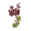
TitleStructure of the human L-type voltage-gated calcium channel Cav1.2 complexed with L-leucine (CABAD Map 2)
Map dataStructure of the human L-type voltage-gated calcium channel Cav1.2 complexed with L-leucine (CABAD Map 2)
Sample
  • Cell: Ternary complex of human CaV alpha1C with rabbit CaV alpha2delta-1 and rabbit CaV beta3
    • Complex: Human CaV alpha1C
      • Protein or peptide: x 1 types
    • Complex: Rabbit CaV alpha2delta-1
      • Protein or peptide: x 1 types
    • Complex: Rabbit CaV beta3
      • Protein or peptide: x 1 types
  • Ligand: x 8 types
Keywordsvoltage-gated calcium channel / CaV alpha2delta / drug binding / gabapentinoid / MEMBRANE PROTEIN
Function / homology
Function and homology information


positive regulation of high voltage-gated calcium channel activity / calcium ion transmembrane transport via high voltage-gated calcium channel / L-type voltage-gated calcium channel complex / calcium channel regulator activity / calcium ion import across plasma membrane / voltage-gated calcium channel activity / T-tubule / calcium channel activity / metal ion binding / cytoplasm
Similarity search - Function
Voltage-dependent calcium channel, L-type, beta-3 subunit / Voltage-dependent L-type calcium channel subunit beta-3, SH3 domain / Voltage-dependent L-type calcium channel subunit beta-1-4, N-terminal A domain / Voltage-dependent calcium channel, L-type, beta subunit / Voltage gated calcium channel subunit beta domain 4Aa N terminal / VWA N-terminal / Voltage-dependent calcium channel, alpha-2/delta subunit, conserved region / VWA N-terminal / Neuronal voltage-dependent calcium channel alpha 2acd / Guanylate kinase/L-type calcium channel beta subunit ...Voltage-dependent calcium channel, L-type, beta-3 subunit / Voltage-dependent L-type calcium channel subunit beta-3, SH3 domain / Voltage-dependent L-type calcium channel subunit beta-1-4, N-terminal A domain / Voltage-dependent calcium channel, L-type, beta subunit / Voltage gated calcium channel subunit beta domain 4Aa N terminal / VWA N-terminal / Voltage-dependent calcium channel, alpha-2/delta subunit, conserved region / VWA N-terminal / Neuronal voltage-dependent calcium channel alpha 2acd / Guanylate kinase/L-type calcium channel beta subunit / Guanylate kinase / Guanylate kinase homologues. / von Willebrand factor type A domain / von Willebrand factor (vWF) type A domain / VWFA domain profile. / von Willebrand factor, type A / von Willebrand factor A-like domain superfamily / SH3-like domain superfamily / Src homology 3 (SH3) domain profile. / SH3 domain / P-loop containing nucleoside triphosphate hydrolase
Similarity search - Domain/homology
Voltage-dependent calcium channel subunit alpha-2/delta-1 / Voltage-dependent L-type calcium channel subunit beta-3 / Isoform 20 of Voltage-dependent L-type calcium channel subunit alpha-1C
Similarity search - Component
Biological speciesHomo sapiens (human) / Oryctolagus cuniculus (rabbit)
Methodsingle particle reconstruction / cryo EM / Resolution: 3.3 Å
AuthorsChen Z / Mondal A / Abderemane-Ali F / Minor DL
Funding support United States, 2 items
OrganizationGrant numberCountry
National Institutes of Health/National Heart, Lung, and Blood Institute (NIH/NHLBI)HL080050 United States
National Institutes of Health/National Institute on Deafness and Other Communication Disorders (NIH/NIDCD)DC007664 United States
CitationJournal: Nature / Year: 2023
Title: EMC chaperone-Ca structure reveals an ion channel assembly intermediate.
Authors: Zhou Chen / Abhisek Mondal / Fayal Abderemane-Ali / Seil Jang / Sangeeta Niranjan / José L Montaño / Balyn W Zaro / Daniel L Minor /
Abstract: Voltage-gated ion channels (VGICs) comprise multiple structural units, the assembly of which is required for function. Structural understanding of how VGIC subunits assemble and whether chaperone ...Voltage-gated ion channels (VGICs) comprise multiple structural units, the assembly of which is required for function. Structural understanding of how VGIC subunits assemble and whether chaperone proteins are required is lacking. High-voltage-activated calcium channels (Cas) are paradigmatic multisubunit VGICs whose function and trafficking are powerfully shaped by interactions between pore-forming Ca1 or Ca2 Caα (ref. ), and the auxiliary Caβ and Caαδ subunits. Here we present cryo-electron microscopy structures of human brain and cardiac Ca1.2 bound with Caβ to a chaperone-the endoplasmic reticulum membrane protein complex (EMC)-and of the assembled Ca1.2-Caβ-Caαδ-1 channel. These structures provide a view of an EMC-client complex and define EMC sites-the transmembrane (TM) and cytoplasmic (Cyto) docks; interaction between these sites and the client channel causes partial extraction of a pore subunit and splays open the Caαδ-interaction site. The structures identify the Caαδ-binding site for gabapentinoid anti-pain and anti-anxiety drugs, show that EMC and Caαδ interactions with the channel are mutually exclusive, and indicate that EMC-to-Caαδ hand-off involves a divalent ion-dependent step and Ca1.2 element ordering. Disruption of the EMC-Ca complex compromises Ca function, suggesting that the EMC functions as a channel holdase that facilitates channel assembly. Together, the structures reveal a Ca assembly intermediate and EMC client-binding sites that could have wide-ranging implications for the biogenesis of VGICs and other membrane proteins.
History
DepositionOct 3, 2022-
Header (metadata) releaseMay 24, 2023-
Map releaseMay 24, 2023-
UpdateJul 26, 2023-
Current statusJul 26, 2023Processing site: RCSB / Status: Released

-
Structure visualization

Supplemental images

Downloads & links

-
Map

FileDownload / File: emd_28375.map.gz / Format: CCP4 / Size: 325 MB / Type: IMAGE STORED AS FLOATING POINT NUMBER (4 BYTES)
AnnotationStructure of the human L-type voltage-gated calcium channel Cav1.2 complexed with L-leucine (CABAD Map 2)
Voxel sizeX=Y=Z: 0.8466 Å
Density
Contour LevelBy AUTHOR: 6.4
Minimum - Maximum-13.172898999999999 - 35.80742
Average (Standard dev.)-0.061263837 (±0.915198)
SymmetrySpace group: 1
Details

EMDB XML:

Map geometry
Axis orderZYX
Origin000
Dimensions440440440
Spacing440440440
CellA=B=C: 372.504 Å
α=β=γ: 90.0 °

-
Supplemental data

-
Sample components

+
Entire : Ternary complex of human CaV alpha1C with rabbit CaV alpha2delta-...

EntireName: Ternary complex of human CaV alpha1C with rabbit CaV alpha2delta-1 and rabbit CaV beta3
Components
  • Cell: Ternary complex of human CaV alpha1C with rabbit CaV alpha2delta-1 and rabbit CaV beta3
    • Complex: Human CaV alpha1C
      • Protein or peptide: Isoform 20 of Voltage-dependent L-type calcium channel subunit alpha-1C
    • Complex: Rabbit CaV alpha2delta-1
      • Protein or peptide: Voltage-dependent calcium channel subunit alpha-2/delta-1Voltage-gated calcium channel
    • Complex: Rabbit CaV beta3
      • Protein or peptide: Voltage-dependent L-type calcium channel subunit beta-3
  • Ligand: 2-acetamido-2-deoxy-beta-D-glucopyranose
  • Ligand: CALCIUM IONCalcium
  • Ligand: SODIUM IONSodium
  • Ligand: LEUCINE
  • Ligand: CHOLESTEROL
  • Ligand: (2R)-3-{[(R)-(2-aminoethoxy)(hydroxy)phosphoryl]oxy}-2-(dodecanoyloxy)propyl dodecanoate
  • Ligand: (2R)-3-{[(R)-(2-aminoethoxy)(hydroxy)phosphoryl]oxy}-2-(dodecanoyloxy)propyl heptadecanoate
  • Ligand: water

+
Supramolecule #1: Ternary complex of human CaV alpha1C with rabbit CaV alpha2delta-...

SupramoleculeName: Ternary complex of human CaV alpha1C with rabbit CaV alpha2delta-1 and rabbit CaV beta3
type: cell / ID: 1 / Parent: 0 / Macromolecule list: #1-#3
Source (natural)Organism: Homo sapiens (human)

+
Supramolecule #2: Human CaV alpha1C

SupramoleculeName: Human CaV alpha1C / type: complex / ID: 2 / Parent: 1 / Macromolecule list: #2
Source (natural)Organism: Homo sapiens (human)
Molecular weightTheoretical: 187 KDa

+
Supramolecule #3: Rabbit CaV alpha2delta-1

SupramoleculeName: Rabbit CaV alpha2delta-1 / type: complex / ID: 3 / Parent: 1 / Macromolecule list: #1
Source (natural)Organism: Oryctolagus cuniculus (rabbit)
Molecular weightTheoretical: 125 KDa

+
Supramolecule #4: Rabbit CaV beta3

SupramoleculeName: Rabbit CaV beta3 / type: complex / ID: 4 / Parent: 1 / Macromolecule list: #3
Source (natural)Organism: Oryctolagus cuniculus (rabbit)
Molecular weightTheoretical: 100 KDa

+
Macromolecule #1: Voltage-dependent calcium channel subunit alpha-2/delta-1

MacromoleculeName: Voltage-dependent calcium channel subunit alpha-2/delta-1
type: protein_or_peptide / ID: 1 / Number of copies: 1 / Enantiomer: LEVO
Source (natural)Organism: Oryctolagus cuniculus (rabbit)
Molecular weightTheoretical: 118.941695 KDa
Recombinant expressionOrganism: Homo sapiens (human)
SequenceString: FPSAVTIKSW VDKMQEDLVT LAKTASGVHQ LVDIYEKYQD LYTVEPNNAR QLVEIAARDI EKLLSNRSKA LVRLALEAEK VQAAHQWRE DFASNEVVYY NAKDDLDPEK NDSEPGSQRI KPVFIDDANF RRQVSYQHAA VHIPTDIYEG STIVLNELNW T SALDDVFK ...String:
FPSAVTIKSW VDKMQEDLVT LAKTASGVHQ LVDIYEKYQD LYTVEPNNAR QLVEIAARDI EKLLSNRSKA LVRLALEAEK VQAAHQWRE DFASNEVVYY NAKDDLDPEK NDSEPGSQRI KPVFIDDANF RRQVSYQHAA VHIPTDIYEG STIVLNELNW T SALDDVFK KNREEDPSLL WQVFGSATGL ARYYPASPWV DNSRTPNKID LYDVRRRPWY IQGAASPKDM LILVDVSGSV SG LTLKLIR TSVSEMLETL SDDDFVNVAS FNSNAQDVSC FQHLVQANVR NKKVLKDAVN NITAKGITDY KKGFSFAFEQ LLN YNVSRA NCNKIIMLFT DGGEERAQEI FAKYNKDKKV RVFTFSVGQH NYDRGPIQWM ACENKGYYYE IPSIGAIRIN TQEY LDVLG RPMVLAGDKA KQVQWTNVYL DALELGLVIT GTLPVFNITG QFENKTNLKN QLILGVMGVD VSLEDIKRLT PRFTL CPNG YYFAIDPNGY VLLHPNLQPK PIGVGIPTIN LRKRRPNVQN PKSQEPVTLD FLDAELENDI KVEIRNKMID GESGEK TFR TLVKSQDERY IDKGNRTYTW TPVNGTDYSL ALVLPTYSFY YIKAKIEETI TQARYSETLK PDNFEESGYT FLAPRDY CS DLKPSDNNTE FLLNFNEFID RKTPNNPSCN TDLINRVLLD AGFTNELVQN YWSKQKNIKG VKARFVVTDG GITRVYPK E AGENWQENPE TYEDSFYKRS LDNDNYVFTA PYFNKSGPGA YESGIMVSKA VEIYIQGKLL KPAVVGIKID VNSWIENFT KTSIRDPCAG PVCDCKRNSD VMDCVILDDG GFLLMANHDD YTNQIGRFFG EIDPSLMRHL VNISVYAFNK SYDYQSVCEP GAAPKQGAG HRSAYVPSIA DILQIGWWAT AAAWSILQQF LLSLTFPRLL EAADMEDDDF TASMSKQSCI TEQTQYFFDN D SKSFSGVL DCGNCSRIFH VEKLMNTNLI FIMVESKGTC PCDTRLLIQA EQTSDGPDPC DMVKQPRYRK GPDVCFDNNV LE DYTDCGG VSG

UniProtKB: Voltage-dependent calcium channel subunit alpha-2/delta-1

+
Macromolecule #2: Isoform 20 of Voltage-dependent L-type calcium channel subunit al...

MacromoleculeName: Isoform 20 of Voltage-dependent L-type calcium channel subunit alpha-1C
type: protein_or_peptide / ID: 2 / Number of copies: 1 / Enantiomer: LEVO
Source (natural)Organism: Homo sapiens (human)
Molecular weightTheoretical: 170.774938 KDa
Recombinant expressionOrganism: Homo sapiens (human)
SequenceString: PIRRACISIV EWKPFEIIIL LTIFANCVAL AIYIPFPEDD SNATNSNLER VEYLFLIIFT VEAFLKVIAY GLLFHPNAYL RNGWNLLDF IIVVVGLFSA ILEQATKADG ANALGGKGAG FDVKALRAFR VLRPLRLVSG VPSLQVVLNS IIKAMVPLLH I ALLVLFVI ...String:
PIRRACISIV EWKPFEIIIL LTIFANCVAL AIYIPFPEDD SNATNSNLER VEYLFLIIFT VEAFLKVIAY GLLFHPNAYL RNGWNLLDF IIVVVGLFSA ILEQATKADG ANALGGKGAG FDVKALRAFR VLRPLRLVSG VPSLQVVLNS IIKAMVPLLH I ALLVLFVI IIYAIIGLEL FMGKMHKTCY NQEGIADVPA EDDPSPCALE TGHGRQCQNG TVCKPGWDGP KHGITNFDNF AF AMLTVFQ CITMEGWTDV LYWVNDAVGR DWPWIYFVTL IIIGSFFVLN LVLGVLSGEF SKEREKAKAR GDFQKLREKQ QLE EDLKGY LDWITQAEDI DPENEDEGMD EEKPRNMSMP TSETESVNTE NVAGGDIEGE NCGARLAHRI SKSKFSRYWR RWNR FCRRK CRAAVKSNVF YWLVIFLVFL NTLTIASEHY NQPNWLTEVQ DTANKALLAL FTAEMLLKMY SLGLQAYFVS LFNRF DCFV VCGGILETIL VETKIMSPLG ISVLRCVRLL RIFKITRYWN SLSNLVASLL NSVRSIASLL LLLFLFIIIF SLLGMQ LFG GKFNFDEMQT RRSTFDNFPQ SLLTVFQILT GEDWNSVMYD GIMAYGGPSF PGMLVCIYFI ILFICGNYIL LNVFLAI AV DNLADAESLT SAQKEEEEEK ERKKLARTAS PEKKQELVEK PAVGESKEEK IELKSITADG ESPPATKINM DDLQPNEN E DKSPYPNPET TGEEDEEEPE MPVGPRPRPL SELHLKEKAV PMPEASAFFI FSSNNRFRLQ CHRIVNDTIF TNLILFFIL LSSISLAAED PVQHTSFRNH ILGNADYVFT SIFTLEIILK MTAYGAFLHK GSFCRNYFNI LDLLVVSVSL ISFGIQSSAI NVVKILRVL RVLRPLRAIN RAKGLKHVVQ CVFVAIRTIG NIVIVTTLLQ FMFACIGVQL FKGKLYTCSD SSKQTEAECK G NYITYKDG EVDHPIIQPR SWENSKFDFD NVLAAMMALF TVSTFEGWPE LLYRSIDSHT EDKGPIYNYR VEISIFFIIY II IIAFFMM NIFVGFVIVT FQEQGEQEYK NCELDKNQRQ CVEYALKARP LRRYIPKNQH QYKVWYVVNS TYFEYLMFVL ILL NTICLA MQHYGQSCLF KIAMNILNML FTGLFTVEMI LKLIAFKPKH YFCDAWNTFD ALIVVGSIVD IAITEVNPAE HTQC SPSMN AEENSRISIT FFRLFRVMRL VKLLSRGEGI RTLLWTFIKS FQALPYVALL IVMLFFIYAV IGMQVFGKIA LNDTT EINR NNNFQTFPQA VLLLFRCATG EAWQDIMLAC MPGKKCAPES EPSNSTEGET PCGSSFAVFY FISFYMLCAF LIINLF VAV IMDNFDYLTR DWSILGPHHL DEFKRIWAEY DPEAKGRIKH LDVVTLLRRI QPPLGFGKLC PHRVACKRLV SMNMPLN SD GTVMFNATLF ALVRTALRIK TEGNLEQANE ELRAIIKKIW KRTSMKLLDQ VVPPAGD

UniProtKB: Isoform 20 of Voltage-dependent L-type calcium channel subunit alpha-1C

+
Macromolecule #3: Voltage-dependent L-type calcium channel subunit beta-3

MacromoleculeName: Voltage-dependent L-type calcium channel subunit beta-3
type: protein_or_peptide / ID: 3 / Number of copies: 1 / Enantiomer: LEVO
Source (natural)Organism: Oryctolagus cuniculus (rabbit)
Molecular weightTheoretical: 21.667154 KDa
Recombinant expressionOrganism: Homo sapiens (human)
SequenceString:
YDVVPSMRPV VLVGPSLKGY EVTDMMQKAL FDFLKHRFDG RISITRVTAD LSLAKRSVLN NPGKRTIIER SSARSSIAEV QSEIERIFE LAKSLQLVVL DADTINHPAQ LAKTSLAPII VFVKVSSPKV LQRLIRSRGK SQMKHLTVQM MAYDKLVQCP P ESFDVILD ENQLEDACEH LAEYLEVYWR ATH

UniProtKB: Voltage-dependent L-type calcium channel subunit beta-3

+
Macromolecule #6: 2-acetamido-2-deoxy-beta-D-glucopyranose

MacromoleculeName: 2-acetamido-2-deoxy-beta-D-glucopyranose / type: ligand / ID: 6 / Number of copies: 5 / Formula: NAG
Molecular weightTheoretical: 221.208 Da
Chemical component information

ChemComp-NAG:
2-acetamido-2-deoxy-beta-D-glucopyranose / N-Acetylglucosamine

+
Macromolecule #7: CALCIUM ION

MacromoleculeName: CALCIUM ION / type: ligand / ID: 7 / Number of copies: 3 / Formula: CA
Molecular weightTheoretical: 40.078 Da

+
Macromolecule #8: SODIUM ION

MacromoleculeName: SODIUM ION / type: ligand / ID: 8 / Number of copies: 1
Molecular weightTheoretical: 22.99 Da

+
Macromolecule #9: LEUCINE

MacromoleculeName: LEUCINE / type: ligand / ID: 9 / Number of copies: 1 / Formula: LEU
Molecular weightTheoretical: 131.173 Da
Chemical component information

ChemComp-LEU:
LEUCINE / Leucine

+
Macromolecule #10: CHOLESTEROL

MacromoleculeName: CHOLESTEROL / type: ligand / ID: 10 / Number of copies: 3 / Formula: CLR
Molecular weightTheoretical: 386.654 Da
Chemical component information

ChemComp-CLR:
CHOLESTEROL / Cholesterol

+
Macromolecule #11: (2R)-3-{[(R)-(2-aminoethoxy)(hydroxy)phosphoryl]oxy}-2-(dodecanoy...

MacromoleculeName: (2R)-3-{[(R)-(2-aminoethoxy)(hydroxy)phosphoryl]oxy}-2-(dodecanoyloxy)propyl dodecanoate
type: ligand / ID: 11 / Number of copies: 1 / Formula: WNZ
Molecular weightTheoretical: 579.746 Da
Chemical component information

ChemComp-WNZ:
(2R)-3-{[(R)-(2-aminoethoxy)(hydroxy)phosphoryl]oxy}-2-(dodecanoyloxy)propyl dodecanoate

+
Macromolecule #12: (2R)-3-{[(R)-(2-aminoethoxy)(hydroxy)phosphoryl]oxy}-2-(dodecanoy...

MacromoleculeName: (2R)-3-{[(R)-(2-aminoethoxy)(hydroxy)phosphoryl]oxy}-2-(dodecanoyloxy)propyl heptadecanoate
type: ligand / ID: 12 / Number of copies: 1 / Formula: WO9
Molecular weightTheoretical: 649.879 Da
Chemical component information

ChemComp-WO9:
(2R)-3-{[(R)-(2-aminoethoxy)(hydroxy)phosphoryl]oxy}-2-(dodecanoyloxy)propyl heptadecanoate

+
Macromolecule #13: water

MacromoleculeName: water / type: ligand / ID: 13 / Number of copies: 3 / Formula: HOH
Molecular weightTheoretical: 18.015 Da
Chemical component information

ChemComp-HOH:
WATER / Water

-
Experimental details

-
Structure determination

Methodcryo EM
Processingsingle particle reconstruction
Aggregation stateparticle

-
Sample preparation

Concentration2.7 mg/mL
BufferpH: 8
GridModel: Quantifoil R1.2/1.3 / Material: GOLD / Mesh: 300 / Support film - Material: CARBON / Support film - topology: HOLEY
VitrificationCryogen name: ETHANE / Chamber humidity: 100 % / Chamber temperature: 277 K / Instrument: FEI VITROBOT MARK IV

-
Electron microscopy

MicroscopeFEI TITAN KRIOS
Electron beamAcceleration voltage: 300 kV / Electron source: FIELD EMISSION GUN
Electron opticsIllumination mode: FLOOD BEAM / Imaging mode: BRIGHT FIELDBright-field microscopy / Cs: 2.7 mm / Nominal defocus max: 1.7 µm / Nominal defocus min: 0.9 µm / Nominal magnification: 105000
Image recordingFilm or detector model: GATAN K3 (6k x 4k) / Average electron dose: 46.0 e/Å2
Experimental equipment
Model: Titan Krios / Image courtesy: FEI Company

-
Image processing

Startup modelType of model: PDB ENTRY
PDB model - PDB ID:
Initial angle assignmentType: MAXIMUM LIKELIHOOD
Final angle assignmentType: MAXIMUM LIKELIHOOD
Final reconstructionApplied symmetry - Point group: C1 (asymmetric) / Resolution.type: BY AUTHOR / Resolution: 3.3 Å / Resolution method: FSC 0.143 CUT-OFF / Number images used: 269802

+
About Yorodumi

-
News

-
Feb 9, 2022. New format data for meta-information of EMDB entries

New format data for meta-information of EMDB entries

  • Version 3 of the EMDB header file is now the official format.
  • The previous official version 1.9 will be removed from the archive.

Related info.:EMDB header

External links:wwPDB to switch to version 3 of the EMDB data model

-
Aug 12, 2020. Covid-19 info

Covid-19 info

URL: https://pdbj.org/emnavi/covid19.php

New page: Covid-19 featured information page in EM Navigator.

Related info.:Covid-19 info / Mar 5, 2020. Novel coronavirus structure data

+
Mar 5, 2020. Novel coronavirus structure data

Novel coronavirus structure data

Related info.:Yorodumi Speices / Aug 12, 2020. Covid-19 info

External links:COVID-19 featured content - PDBj / Molecule of the Month (242):Coronavirus Proteases

+
Jan 31, 2019. EMDB accession codes are about to change! (news from PDBe EMDB page)

EMDB accession codes are about to change! (news from PDBe EMDB page)

  • The allocation of 4 digits for EMDB accession codes will soon come to an end. Whilst these codes will remain in use, new EMDB accession codes will include an additional digit and will expand incrementally as the available range of codes is exhausted. The current 4-digit format prefixed with “EMD-” (i.e. EMD-XXXX) will advance to a 5-digit format (i.e. EMD-XXXXX), and so on. It is currently estimated that the 4-digit codes will be depleted around Spring 2019, at which point the 5-digit format will come into force.
  • The EM Navigator/Yorodumi systems omit the EMD- prefix.

Related info.:Q: What is EMD? / ID/Accession-code notation in Yorodumi/EM Navigator

External links:EMDB Accession Codes are Changing Soon! / Contact to PDBj

+
Jul 12, 2017. Major update of PDB

Major update of PDB

  • wwPDB released updated PDB data conforming to the new PDBx/mmCIF dictionary.
  • This is a major update changing the version number from 4 to 5, and with Remediation, in which all the entries are updated.
  • In this update, many items about electron microscopy experimental information are reorganized (e.g. em_software).
  • Now, EM Navigator and Yorodumi are based on the updated data.

External links:wwPDB Remediation / Enriched Model Files Conforming to OneDep Data Standards Now Available in the PDB FTP Archive

-
Yorodumi

Thousand views of thousand structures

  • Yorodumi is a browser for structure data from EMDB, PDB, SASBDB, etc.
  • This page is also the successor to EM Navigator detail page, and also detail information page/front-end page for Omokage search.
  • The word "yorodu" (or yorozu) is an old Japanese word meaning "ten thousand". "mi" (miru) is to see.

Related info.:EMDB / PDB / SASBDB / Comparison of 3 databanks / Yorodumi Search / Aug 31, 2016. New EM Navigator & Yorodumi / Yorodumi Papers / Jmol/JSmol / Function and homology information / Changes in new EM Navigator and Yorodumi

Read more