 Movie
Movie Controller

[English] 日本語
 Yorodumi
- EMDB-25881: Cryo-EM structure of PilA-N and PilA-C from Geobacter sulfurreducens -
+ Open data

- Basic information
| Entry | Database: EMDB / ID: EMD-25881 | |||||||||
|---|---|---|---|---|---|---|---|---|---|---|
| Title | Cryo-EM structure of PilA-N and PilA-C from Geobacter sulfurreducens | |||||||||
 Map data Map data | Cryo-EM structure of PilA-N and PilA-C from Geobacter sulfurreducens | |||||||||
 Sample Sample |
| |||||||||
 Keywords Keywords |  helical symmetry / filament / pili / type iv pili / pseudo pili / PROTEIN FIBRIL helical symmetry / filament / pili / type iv pili / pseudo pili / PROTEIN FIBRIL | |||||||||
| Function / homology |  Function and homology informationpilus assembly / protein secretion by the type II secretion system / type II protein secretion system complex / membrane Function and homology informationpilus assembly / protein secretion by the type II secretion system / type II protein secretion system complex / membraneSimilarity search - Function | |||||||||
| Biological species |  Geobacter sulfurreducens (bacteria) Geobacter sulfurreducens (bacteria) | |||||||||
| Method | helical reconstruction / cryo EM / Resolution: 4.1 Å | |||||||||
 Authors Authors | Wang F / Mustafa K | |||||||||
| Funding support |  United States, 2 items United States, 2 items
| |||||||||
 Citation Citation | Journal: Nat Microbiol / Year: 2022 Title: Cryo-EM structure of an extracellular Geobacter OmcE cytochrome filament reveals tetrahaem packing. Authors: Fengbin Wang / Khawla Mustafa / Victor Suciu / Komal Joshi / Chi H Chan / Sol Choi / Zhangli Su / Dong Si / Allon I Hochbaum / Edward H Egelman / Daniel R Bond / Abstract: Electrically conductive appendages from the anaerobic bacterium Geobacter sulfurreducens were first observed two decades ago, with genetic and biochemical data suggesting that conductive fibres were ...Electrically conductive appendages from the anaerobic bacterium Geobacter sulfurreducens were first observed two decades ago, with genetic and biochemical data suggesting that conductive fibres were type IV pili. Recently, an extracellular conductive filament of G. sulfurreducens was found to contain polymerized c-type cytochrome OmcS subunits, not pilin subunits. Here we report that G. sulfurreducens also produces a second, thinner appendage comprised of cytochrome OmcE subunits and solve its structure using cryo-electron microscopy at ~4.3 Å resolution. Although OmcE and OmcS subunits have no overall sequence or structural similarities, upon polymerization both form filaments that share a conserved haem packing arrangement in which haems are coordinated by histidines in adjacent subunits. Unlike OmcS filaments, OmcE filaments are highly glycosylated. In extracellular fractions from G. sulfurreducens, we detected type IV pili comprising PilA-N and -C chains, along with abundant B-DNA. OmcE is the second cytochrome filament to be characterized using structural and biophysical methods. We propose that there is a broad class of conductive bacterial appendages with conserved haem packing (rather than sequence homology) that enable long-distance electron transport to chemicals or other microbial cells. | |||||||||
| History |
|
- Structure visualization
| Movie |
Movie viewer |
|---|---|
| Structure viewer | EM map: SurfViewMolmilJmol/JSmol |
| Supplemental images |

- Downloads & links
-EMDB archive
| Map data | emd_25881.map.gz | 8.6 MB | EMDB map data format | |
|---|---|---|---|---|
| Header (meta data) | emd-25881-v30.xmlemd-25881.xml | 10.8 KB 10.8 KB | Display Display | EMDB header |
| Images | 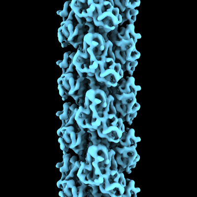 emd_25881.png emd_25881.png | 129.2 KB | ||
| Filedesc metadata | emd-25881.cif.gz | 4.9 KB | ||
| Archive directory |  http://ftp.pdbj.org/pub/emdb/structures/EMD-25881ftp://ftp.pdbj.org/pub/emdb/structures/EMD-25881 http://ftp.pdbj.org/pub/emdb/structures/EMD-25881ftp://ftp.pdbj.org/pub/emdb/structures/EMD-25881 | HTTPS FTP |
-Related structure data
| Related structure data |  7tggMC  7tfsC M: atomic model generated by this map C: citing same article ( |
|---|---|
| Similar structure data |


-Links
| EMDB pages | EMDB (EBI/PDBe) / EMDataResource |
|---|---|
| Related items in Molecule of the Month |

-Map
| File | Download / File: emd_25881.map.gz / Format: CCP4 / Size: 125 MB / Type: IMAGE STORED AS FLOATING POINT NUMBER (4 BYTES) | ||||||||||||||||||||||||||||||||||||||||||||||||||||||||||||
|---|---|---|---|---|---|---|---|---|---|---|---|---|---|---|---|---|---|---|---|---|---|---|---|---|---|---|---|---|---|---|---|---|---|---|---|---|---|---|---|---|---|---|---|---|---|---|---|---|---|---|---|---|---|---|---|---|---|---|---|---|---|
| Annotation | Cryo-EM structure of PilA-N and PilA-C from Geobacter sulfurreducens | ||||||||||||||||||||||||||||||||||||||||||||||||||||||||||||
| Voxel size | X=Y=Z: 1.08 Å | ||||||||||||||||||||||||||||||||||||||||||||||||||||||||||||
| Density |
| ||||||||||||||||||||||||||||||||||||||||||||||||||||||||||||
| Symmetry | Space group: 1 | ||||||||||||||||||||||||||||||||||||||||||||||||||||||||||||
| Details | EMDB XML:
CCP4 map header:
| ||||||||||||||||||||||||||||||||||||||||||||||||||||||||||||
-Supplemental data
- Sample components
-Entire : Filament of PilA-N and PilA-C proteins
| Entire | Name: Filament of PilA-N and PilA-C proteins |
|---|---|
| Components |
|
-Supramolecule #1: Filament of PilA-N and PilA-C proteins
| Supramolecule | Name: Filament of PilA-N and PilA-C proteins / type: complex / ID: 1 / Parent: 0 / Macromolecule list: all |
|---|---|
| Source (natural) | Organism: Geobacter sulfurreducens (bacteria) / Strain: ATCC 51573 / DSM 12127 / PCA |
-Macromolecule #1: Geopilin domain 1 protein
| Macromolecule | Name: Geopilin domain 1 protein / type: protein_or_peptide / ID: 1 / Number of copies: 1 / Enantiomer: LEVO |
|---|---|
| Source (natural) | Organism: Geobacter sulfurreducens (bacteria) / Strain: ATCC 51573 / DSM 12127 / PCA |
| Molecular weight | Theoretical: 10.006582 KDa |
| Sequence | String: MANYPHTPTQ AAKRRKETLM LQKLRNRKGF TLIELLIVVA IIGILAAIAI PQFSAYRVKA YNSAASSDLR NLKTALESAF ADDQTYPPE S UniProtKB: Geopilin domain 1 protein |
-Macromolecule #2: Geopilin domain 2 protein
| Macromolecule | Name: Geopilin domain 2 protein / type: protein_or_peptide / ID: 2 / Number of copies: 1 / Enantiomer: LEVO |
|---|---|
| Source (natural) | Organism: Geobacter sulfurreducens (bacteria) / Strain: ATCC 51573 / DSM 12127 / PCA |
| Molecular weight | Theoretical: 13.087543 KDa |
| Sequence | String: MKKIITIVAM LLAMQGIAIA AGKIPTTTMG GKDFTFKPST NVSVSYFTTN GATSTAGTVN TDYAVNTKNS SGNRVFTSTN NTSNIWYIE NDAWKGKAVS DSDVTALGTG DVGKSDFSGT EWKSQ UniProtKB: Geopilin domain 2 protein |
-Experimental details
-Structure determination
| Method | cryo EM |
|---|---|
 Processing Processing | helical reconstruction |
| Aggregation state | filament |
-Sample preparation
| Buffer | pH: 6 |
|---|---|
| Vitrification | Cryogen name: ETHANE |
- Electron microscopy
| Microscope | FEI TITAN KRIOS |
|---|---|
| Electron beam | Acceleration voltage: 300 kV / Electron source: FIELD EMISSION GUN |
| Electron optics | Illumination mode: FLOOD BEAM / Imaging mode: BRIGHT FIELDBright-field microscopy / Nominal defocus max: 3.0 µm / Nominal defocus min: 1.0 µm |
| Image recording | Film or detector model: GATAN K3 (6k x 4k) / Average electron dose: 50.0 e/Å2 |
| Experimental equipment |  Model: Titan Krios / Image courtesy: FEI Company |
-Image processing
| Startup model | Type of model: NONE |
|---|---|
| Final angle assignment | Type: NOT APPLICABLE |
| Final reconstruction | Applied symmetry - Helical parameters - Δz: 10.4 Å Applied symmetry - Helical parameters - Δ&Phi: 89.1 ° Applied symmetry - Helical parameters - Axial symmetry: C1 (asymmetric) Resolution.type: BY AUTHOR / Resolution: 4.1 Å / Resolution method: FSC 0.143 CUT-OFF / Number images used: 112011 |
