 Movie
Movie Controller

[English] 日本語
 Yorodumi
- EMDB-25581: cryoEM reconstruction of the HIV gp140 in complex with the extrac... -
+ Open data

- Basic information
| Entry | Database: EMDB / ID: EMD-25581 | |||||||||
|---|---|---|---|---|---|---|---|---|---|---|
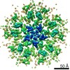
| Title | cryoEM reconstruction of the HIV gp140 in complex with the extracellular domains of CD4 and the adnectin domain of Combinectin. The gp140 and CD4 coordinates from entry 6EDU were rigid body fitted to the EM map along withe the crystal structure of CD4+adnectin | |||||||||
 Map data Map data | ||||||||||
 Sample Sample |
| |||||||||
| Function / homology |  Function and homology information Function and homology informationhelper T cell enhancement of adaptive immune response /  interleukin-16 binding / interleukin-16 receptor activity / maintenance of protein location in cell / T cell selection / MHC class II protein binding / cellular response to granulocyte macrophage colony-stimulating factor stimulus / interleukin-15-mediated signaling pathway / positive regulation of monocyte differentiation / Nef Mediated CD4 Down-regulation ...helper T cell enhancement of adaptive immune response / interleukin-16 binding / interleukin-16 receptor activity / maintenance of protein location in cell / T cell selection / MHC class II protein binding / cellular response to granulocyte macrophage colony-stimulating factor stimulus / interleukin-15-mediated signaling pathway / positive regulation of monocyte differentiation / Nef Mediated CD4 Down-regulation / Alpha-defensins / positive regulation of kinase activity / regulation of T cell activation / T cell receptor complex / extracellular matrix structural constituent / Other interleukin signaling / enzyme-linked receptor protein signaling pathway / Translocation of ZAP-70 to Immunological synapse / Phosphorylation of CD3 and TCR zeta chains / regulation of calcium ion transport / macrophage differentiation / Generation of second messenger molecules / T cell differentiation / PD-1 signaling / positive regulation of protein kinase activity / Binding and entry of HIV virion / coreceptor activity / positive regulation of calcium-mediated signaling / protein tyrosine kinase binding / positive regulation of interleukin-2 production / T cell activation / Vpu mediated degradation of CD4 / calcium-mediated signaling / clathrin-coated endocytic vesicle membrane / cell surface receptor protein tyrosine kinase signaling pathway / positive regulation of T cell activation / positive regulation of peptidyl-tyrosine phosphorylation / transmembrane signaling receptor activity / Cargo recognition for clathrin-mediated endocytosis / Downstream TCR signaling / virus receptor activity / MHC class II protein complex binding / Clathrin-mediated endocytosis / signaling receptor activity / positive regulation of canonical NF-kappaB signal transduction / defense response to Gram-negative bacterium / adaptive immune response / positive regulation of MAPK cascade / positive regulation of viral entry into host cell / cell surface receptor signaling pathway / positive regulation of ERK1 and ERK2 cascade / early endosome / cell adhesion / immune response / positive regulation of protein phosphorylation / membrane raft / external side of plasma membrane / endoplasmic reticulum lumen / lipid binding / endoplasmic reticulum membrane / protein kinase binding / positive regulation of DNA-templated transcription / enzyme binding / signal transduction / protein homodimerization activity / zinc ion binding / identical protein binding / plasma membrane interleukin-16 binding / interleukin-16 receptor activity / maintenance of protein location in cell / T cell selection / MHC class II protein binding / cellular response to granulocyte macrophage colony-stimulating factor stimulus / interleukin-15-mediated signaling pathway / positive regulation of monocyte differentiation / Nef Mediated CD4 Down-regulation ...helper T cell enhancement of adaptive immune response / interleukin-16 binding / interleukin-16 receptor activity / maintenance of protein location in cell / T cell selection / MHC class II protein binding / cellular response to granulocyte macrophage colony-stimulating factor stimulus / interleukin-15-mediated signaling pathway / positive regulation of monocyte differentiation / Nef Mediated CD4 Down-regulation / Alpha-defensins / positive regulation of kinase activity / regulation of T cell activation / T cell receptor complex / extracellular matrix structural constituent / Other interleukin signaling / enzyme-linked receptor protein signaling pathway / Translocation of ZAP-70 to Immunological synapse / Phosphorylation of CD3 and TCR zeta chains / regulation of calcium ion transport / macrophage differentiation / Generation of second messenger molecules / T cell differentiation / PD-1 signaling / positive regulation of protein kinase activity / Binding and entry of HIV virion / coreceptor activity / positive regulation of calcium-mediated signaling / protein tyrosine kinase binding / positive regulation of interleukin-2 production / T cell activation / Vpu mediated degradation of CD4 / calcium-mediated signaling / clathrin-coated endocytic vesicle membrane / cell surface receptor protein tyrosine kinase signaling pathway / positive regulation of T cell activation / positive regulation of peptidyl-tyrosine phosphorylation / transmembrane signaling receptor activity / Cargo recognition for clathrin-mediated endocytosis / Downstream TCR signaling / virus receptor activity / MHC class II protein complex binding / Clathrin-mediated endocytosis / signaling receptor activity / positive regulation of canonical NF-kappaB signal transduction / defense response to Gram-negative bacterium / adaptive immune response / positive regulation of MAPK cascade / positive regulation of viral entry into host cell / cell surface receptor signaling pathway / positive regulation of ERK1 and ERK2 cascade / early endosome / cell adhesion / immune response / positive regulation of protein phosphorylation / membrane raft / external side of plasma membrane / endoplasmic reticulum lumen / lipid binding / endoplasmic reticulum membrane / protein kinase binding / positive regulation of DNA-templated transcription / enzyme binding / signal transduction / protein homodimerization activity / zinc ion binding / identical protein binding / plasma membraneSimilarity search - Function | |||||||||
| Biological species |  HIV-1 06TG.HT008 (virus) / HIV-1 06TG.HT008 (virus) /  Homo sapiens (human) Homo sapiens (human) | |||||||||
| Method | single particle reconstruction / cryo EM / Resolution: 8.7 Å | |||||||||
 Authors Authors | Concha NO / William SP / Wenzel DL | |||||||||
| Funding support |  United States, 1 items United States, 1 items
| |||||||||
 Citation Citation | Journal: J Mol Biol / Year: 2022 Title: Novel Bent Conformation of CD4 Induced by HIV-1 Inhibitor Indirectly Prevents Productive Viral Attachment. Authors: David Wensel / Shawn Williams / David P Dixon / Paris Ward / Patti McCormick / Nestor Concha / Eugene Stewart / Xuan Hong / Charles Mazzucco / Shreya Pal / Bo Ding / Christoph Fellinger / Mark Krystal /  Abstract: GSK3732394 is a multi-specific biologic inhibitor of HIV entry currently under clinical evaluation. A key component of this molecule is an adnectin (6940_B01) that binds to CD4 and inhibits ...GSK3732394 is a multi-specific biologic inhibitor of HIV entry currently under clinical evaluation. A key component of this molecule is an adnectin (6940_B01) that binds to CD4 and inhibits downstream actions of gp160. Studies were performed to determine the binding site of the adnectin on CD4 and to understand the mechanism of inhibition. Using hydrogen-deuterium exchange with mass spectrometry (HDX), CD4 peptides showed differential rates of deuteration (either enhanced or slowed) in the presence of the adnectin that mapped predominantly to the interface of domains 2 and 3 (D2-D3). In addition, an X-ray crystal structure of an ibalizumab Fab/CD4(D1-D4)/adnectin complex revealed an extensive interface between the adnectin and residues on CD4 domains D2-D4 that stabilize a novel T-shaped CD4 conformation. A cryo-EM map of the gp140/CD4/GSK3732394 complex clearly shows the bent conformation for CD4 while bound to gp140. Mutagenic analyses on CD4 confirmed that amino acid F202 forms a key interaction with the adnectin. In addition, amino acid L151 was shown to be a critical indirect determinant of the specificity for binding to the human CD4 protein over related primate CD4 molecules, as it appears to modulate CD4's flexibility to adopt the adnectin-bound conformation. The significant conformational change of CD4 upon adnectin binding brings the D1 domain of CD4 in proximity to the host cell membrane surface, thereby re-orienting the gp120 binding site in a direction that is inaccessible to incoming virus due to a steric clash between gp160 trimers on the virus surface and the target cell membrane. | |||||||||
| History |
|
- Structure visualization
| Movie |
Movie viewer |
|---|---|
| Structure viewer | EM map: SurfViewMolmilJmol/JSmol |
| Supplemental images |

- Downloads & links
-EMDB archive
| Map data | emd_25581.map.gz | 1.6 MB | EMDB map data format | |
|---|---|---|---|---|
| Header (meta data) | emd-25581-v30.xmlemd-25581.xml | 21.7 KB 21.7 KB | Display Display | EMDB header |
| FSC (resolution estimation) | emd_25581_fsc.xml | 6.5 KB | Display | FSC data file |
| Images |  emd_25581.png emd_25581.png | 65 KB | ||
| Masks | emd_25581_msk_1.map | 22.2 MB | Mask map | |
| Archive directory |  http://ftp.pdbj.org/pub/emdb/structures/EMD-25581ftp://ftp.pdbj.org/pub/emdb/structures/EMD-25581 http://ftp.pdbj.org/pub/emdb/structures/EMD-25581ftp://ftp.pdbj.org/pub/emdb/structures/EMD-25581 | HTTPS FTP |
-Related structure data
| Related structure data |  7t0oMC  7t0rC M: atomic model generated by this map C: citing same article ( |
|---|---|
| Similar structure data |


-Links
| EMDB pages | EMDB (EBI/PDBe) / EMDataResource |
|---|---|
| Related items in Molecule of the Month |


-Map
| File | Download / File: emd_25581.map.gz / Format: CCP4 / Size: 1.8 MB / Type: IMAGE STORED AS FLOATING POINT NUMBER (4 BYTES) | ||||||||||||||||||||||||||||||||||||||||||||||||||||||||||||||||||||
|---|---|---|---|---|---|---|---|---|---|---|---|---|---|---|---|---|---|---|---|---|---|---|---|---|---|---|---|---|---|---|---|---|---|---|---|---|---|---|---|---|---|---|---|---|---|---|---|---|---|---|---|---|---|---|---|---|---|---|---|---|---|---|---|---|---|---|---|---|---|
| Voxel size | X=Y=Z: 2.4624 Å | ||||||||||||||||||||||||||||||||||||||||||||||||||||||||||||||||||||
| Density |
| ||||||||||||||||||||||||||||||||||||||||||||||||||||||||||||||||||||
| Symmetry | Space group: 1 | ||||||||||||||||||||||||||||||||||||||||||||||||||||||||||||||||||||
| Details | EMDB XML:
CCP4 map header:
| ||||||||||||||||||||||||||||||||||||||||||||||||||||||||||||||||||||
-Supplemental data
-Mask #1
| File | emd_25581_msk_1.map | ||||||||||||
|---|---|---|---|---|---|---|---|---|---|---|---|---|---|
| Projections & Slices |
| ||||||||||||
| Density Histograms |
 Z
Z Y
Y X
X


- Sample components
-Entire : Ternary complex of the HIV-1 gp140 trimer in complex with the ext...
| Entire | Name: Ternary complex of the HIV-1 gp140 trimer in complex with the extracellular domains (domains 1-4) of CD4 and an adnectin domain of Combinectin |
|---|---|
| Components |
|
-Supramolecule #1: Ternary complex of the HIV-1 gp140 trimer in complex with the ext...
| Supramolecule | Name: Ternary complex of the HIV-1 gp140 trimer in complex with the extracellular domains (domains 1-4) of CD4 and an adnectin domain of Combinectin type: complex / Chimera: Yes / ID: 1 / Parent: 0 / Macromolecule list: all Details: the gp140 trimer at 0.67 mg/ml was mixed with soluble CD4 first, and then with Combinection in a 1.25:1 ratio. The sample was incubated overnight at 4 degC. The sample was concentrated to ...Details: the gp140 trimer at 0.67 mg/ml was mixed with soluble CD4 first, and then with Combinection in a 1.25:1 ratio. The sample was incubated overnight at 4 degC. The sample was concentrated to 35ul before injecting into a Phenomenex 4.6 x 300 mm 3um bead 500Angstroms pore Yara SEC column equilibrated with 20mM Hepes pH7.5, 200 mM KCl, 5mM MgCl2. The complexes were eluted with a flow rate of 0.2 ml/min and fractions collected every 20 seconds into a Thermo-Fisher UV transparent 96-well plates. The absorbance at 280nm was measured for each fraction using a Spectramax plate scanner. The high-molecular weight complex appeared to be comprised of gp140:CD4:Combinectin in a 1:1:1 ratio. The sample used for cryo-EM studies was the single peak fraction of gp140/CD4/Combinectin (0.076 mg/ml) |
|---|
-Macromolecule #1: BG505 SOSIP.664 gp140
| Macromolecule | Name: BG505 SOSIP.664 gp140 / type: protein_or_peptide / ID: 1 / Number of copies: 3 / Enantiomer: LEVO |
|---|---|
| Source (natural) | Organism: HIV-1 06TG.HT008 (virus) |
| Molecular weight | Theoretical: 72.830398 KDa |
| Recombinant expression | Organism: Homo sapiens (human) |
| Sequence | String: AENLWVTVYY GVPVWKDAET TLFCASDAKA YETEKHNVWA THACVPTDPN PQEIHLENVT EEFNMWKNNM VEQMHTDIIS LWDQSLKPC VKLTPLCVTL NCTNVTNNIT DDMRGELKNC SFNMTTELRD KKQKVYSLFY RLDVVQINEN QGNRSNNSNK E YRLINCNT ...String: AENLWVTVYY GVPVWKDAET TLFCASDAKA YETEKHNVWA THACVPTDPN PQEIHLENVT EEFNMWKNNM VEQMHTDIIS LWDQSLKPC VKLTPLCVTL NCTNVTNNIT DDMRGELKNC SFNMTTELRD KKQKVYSLFY RLDVVQINEN QGNRSNNSNK E YRLINCNT SAITQACPKV SFEPIPIHYC APAGFAILKC KDKKFNGTGP CPSVSTVQCT HGIKPVVSTQ LLLNGSLAEE EV MIRSENI TNNAKNILVQ FNTPVQINCT RPNNNTRKSI RIGPGQAFYA TGDIIGDIRQ AHCNVSKATW NETLGKVVKQ LRK HFGNNT IIRFANSSGG DLEVTTHSFN CGGEFFYCNT SGLFNSTWIS NTSVQGSNST GSNDSITLPC RIKQIINMWQ RIGQ AMYAP PIQGVIRCVS NITGLILTRD GGSTNSTTET FRPGGGDMRD NWRSELYKYK VVKIEPLGVA PTRCKRRVVG RRRRR RAVG IGAVFLGFLG AAGSTMGAAS MTLTVQARNL LSGIVQQQSN LLRAPEAQQH LLKLTVWGIK QLQARVLAVE RYLRDQ QLL GIWGCSGKLI CCTNVPWNSS WSNRNLSEIW DNMTWLQWDK EISNYTQIIY GLLEESQNQQ EKNEQDLLAL DGSGSGG SG HHHHHHHH |
-Macromolecule #2: BG505 SOSIP.664 gp140
| Macromolecule | Name: BG505 SOSIP.664 gp140 / type: protein_or_peptide / ID: 2 / Number of copies: 1 / Enantiomer: LEVO |
|---|---|
| Source (natural) | Organism: HIV-1 06TG.HT008 (virus) |
| Molecular weight | Theoretical: 3.337105 KDa |
| Recombinant expression | Organism: Homo sapiens (human) |
| Sequence | String: (UNK)(UNK)(UNK)(UNK)(UNK)(UNK)(UNK)(UNK)(UNK)(UNK) (UNK)(UNK)(UNK)(UNK)(UNK)(UNK) (UNK)(UNK)(UNK) (UNK)(UNK)(UNK)(UNK)(UNK)(UNK)(UNK)(UNK)(UNK)(UNK) (UNK)(UNK)(UNK) (UNK)(UNK)(UNK)(UNK)(UNK)(UNK) (UNK) |
-Macromolecule #3: BG505 SOSIP.664 gp140
| Macromolecule | Name: BG505 SOSIP.664 gp140 / type: protein_or_peptide / ID: 3 / Number of copies: 2 / Enantiomer: LEVO |
|---|---|
| Source (natural) | Organism: HIV-1 06TG.HT008 (virus) |
| Molecular weight | Theoretical: 3.252 KDa |
| Recombinant expression | Organism: Homo sapiens (human) |
| Sequence | String: (UNK)(UNK)(UNK)(UNK)(UNK)(UNK)(UNK)(UNK)(UNK)(UNK) (UNK)(UNK)(UNK)(UNK)(UNK)(UNK) (UNK)(UNK)(UNK) (UNK)(UNK)(UNK)(UNK)(UNK)(UNK)(UNK)(UNK)(UNK)(UNK) (UNK)(UNK)(UNK) (UNK)(UNK)(UNK)(UNK)(UNK)(UNK) |
-Macromolecule #4: T-cell surface glycoprotein CD4
| Macromolecule | Name: T-cell surface glycoprotein CD4 / type: protein_or_peptide / ID: 4 / Number of copies: 3 / Enantiomer: LEVO |
|---|---|
| Source (natural) | Organism: Homo sapiens (human) |
| Molecular weight | Theoretical: 41.385449 KDa |
| Recombinant expression | Organism:   Spodoptera frugiperda (fall armyworm) Spodoptera frugiperda (fall armyworm) |
| Sequence | String: KKVVLGKKGD TVELTCTASQ KKSIQFHWKN SNQIKILGNQ GSFLTKGPSK LNDRADSRRS LWDQGNFPLI IKNLKIEDSD TYICEVEDQ KEEVQLLVFG LTANSDTHLL QGQSLTLTLE SPPGSSPSVQ CRSPRGKNIQ GGKTLSVSQL ELQDSGTWTC T VLQNQKKV ...String: KKVVLGKKGD TVELTCTASQ KKSIQFHWKN SNQIKILGNQ GSFLTKGPSK LNDRADSRRS LWDQGNFPLI IKNLKIEDSD TYICEVEDQ KEEVQLLVFG LTANSDTHLL QGQSLTLTLE SPPGSSPSVQ CRSPRGKNIQ GGKTLSVSQL ELQDSGTWTC T VLQNQKKV EFKIDIVVLA FQKASSIVYK KEGEQVEFSF PLAFTVEKLT GSGELWWQAE RASSSKSWIT FDLKNKEVSV KR VTQDPKL QMGKKLPLHL TLPQALPQYA GSGNLTLALE AKTGKLHQEV NLVVMRATQL QKNLTCEVWG PTSPKLMLSL KLE NKEAKV SKREKAVWVL NPEAGMWQCL LSDSGQVLLE SNIKVLPTHH HHHH |
-Macromolecule #5: Adnectin
| Macromolecule | Name: Adnectin / type: protein_or_peptide / ID: 5 Details: The native 10th fibronectin type III (10Fn3) domain from human fibronectin was used as the template for construction of novel libraries of Adnectin variants (Wenzel, D., et al. (2017) ...Details: The native 10th fibronectin type III (10Fn3) domain from human fibronectin was used as the template for construction of novel libraries of Adnectin variants (Wenzel, D., et al. (2017) Antimicrobial Agents and Chemotherapy, Vol 61(8), e00508-17, pp 1-19) Number of copies: 3 / Enantiomer: LEVO |
|---|---|
| Source (natural) | Organism: Homo sapiens (human) |
| Molecular weight | Theoretical: 11.959172 KDa |
| Recombinant expression | Organism:  Escherichia coli (E. coli) Escherichia coli (E. coli) |
| Sequence | String: GVSDVPRDLE VVAATPTSLL ISWDAPAVTV HSYHIQYWPL GSYQRYQVFS VPGSKSTATI SGLKPGVEYQ IRVYAETGGA DSDQSMGWI QIGYRTEGDK PSQHHHHHH |
-Macromolecule #6: BG505 SOSIP.664 gp140
| Macromolecule | Name: BG505 SOSIP.664 gp140 / type: protein_or_peptide / ID: 6 / Number of copies: 3 / Enantiomer: LEVO |
|---|---|
| Source (natural) | Organism: HIV-1 06TG.HT008 (virus) |
| Molecular weight | Theoretical: 2.996685 KDa |
| Recombinant expression | Organism: Homo sapiens (human) |
| Sequence | String: (UNK)(UNK)(UNK)(UNK)(UNK)(UNK)(UNK)(UNK)(UNK)(UNK) (UNK)(UNK)(UNK)(UNK)(UNK)(UNK) (UNK)(UNK)(UNK) (UNK)(UNK)(UNK)(UNK)(UNK)(UNK)(UNK)(UNK)(UNK)(UNK) (UNK)(UNK)(UNK) (UNK)(UNK)(UNK) |
-Experimental details
-Structure determination
| Method | cryo EM |
|---|---|
 Processing Processing | single particle reconstruction |
| Aggregation state | particle |
-Sample preparation
| Concentration | 0.08 mg/mL | ||||||||
|---|---|---|---|---|---|---|---|---|---|
| Buffer | pH: 7.5 Component:
| ||||||||
| Grid | Model: Quantifoil R1.2/1.3 / Material: COPPER / Mesh: 400 / Pretreatment - Type: GLOW DISCHARGE / Pretreatment - Atmosphere: AIR / Pretreatment - Pressure: 30.0 kPa Details: glow discharged for 20 sec using a EasyGlo (20mA, 0.3 mBar) | ||||||||
| Vitrification | Cryogen name: ETHANE / Chamber humidity: 100 % / Chamber temperature: 277 K / Instrument: FEI VITROBOT MARK IV | ||||||||
| Details | the gp140/CD4/Combinectin (0.076 mg/ml), 3.5uL were applied to Quantifoil 1.2/1.3 Cu 400 mesh grids that had previously been glow discharged for 20 sec using a EasyGlo (20mA, 0.3 mBar). The grids were plunged into liquid ethane using a Vitrobot plunger (chamber set to 4 degC and 80% RH) immediately after blotting from 2.5 to 4 sec |
- Electron microscopy
| Microscope | FEI TITAN KRIOS |
|---|---|
| Electron beam | Acceleration voltage: 300 kV / Electron source: FIELD EMISSION GUN |
| Electron optics | Illumination mode: FLOOD BEAM / Imaging mode: BRIGHT FIELDBright-field microscopy / Nominal defocus max: 3.4 µm / Nominal defocus min: 1.4000000000000001 µm |
| Specialist optics | Phase plate: OTHER / Spherical aberration corrector: Cs corrector |
| Image recording | Film or detector model: FEI FALCON III (4k x 4k) / Detector mode: COUNTING / Digitization - Dimensions - Width: 4096 pixel / Digitization - Dimensions - Height: 4096 pixel / Digitization - Sampling interval: 11.0 µm / Number real images: 1078 / Average exposure time: 75.7 sec. / Average electron dose: 1.022 e/Å2 |
| Experimental equipment |  Model: Titan Krios / Image courtesy: FEI Company |
-Image processing
| CTF correction | Software - Name: CTFFIND |
|---|---|
| Startup model | Type of model: OTHER / Details: initial model generated with cisTEM |
| Initial angle assignment | Type: MAXIMUM LIKELIHOOD / Software - Name: RELION |
| Final 3D classification | Number classes: 4 / Software - Name: RELION |
| Final angle assignment | Type: MAXIMUM LIKELIHOOD / Software - Name: RELION |
| Final reconstruction | Number classes used: 3 / Applied symmetry - Point group: C3 (3 fold cyclic) / Resolution.type: BY AUTHOR / Resolution: 8.7 Å / Resolution method: FSC 0.143 CUT-OFF / Software - Name: RELION / Number images used: 208485 |
| FSC plot (resolution estimation) |  |
-Atomic model buiding 1
| Refinement | Protocol: RIGID BODY FIT |
|---|---|
| Output model | PDB-7t0o: |
