 Movie
Movie Controller

[English] 日本語
 Yorodumi
- EMDB-24123: Structural basis for enhanced infectivity and immune evasion of S... -
+ Open data

- Basic information
| Entry | Database: EMDB / ID: EMD-24123 | ||||||||||||
|---|---|---|---|---|---|---|---|---|---|---|---|---|---|
| Title | Structural basis for enhanced infectivity and immune evasion of SARS-CoV-2 variants | ||||||||||||
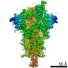
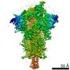
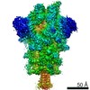
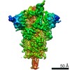
 Map data Map data | overall map | ||||||||||||
 Sample Sample |
| ||||||||||||
| Function / homology |  Function and homology information Function and homology informationMaturation of spike protein / viral translation / Translation of Structural Proteins / Virion Assembly and Release / host cell surface / host extracellular space / suppression by virus of host tetherin activity / Induction of Cell-Cell Fusion / structural constituent of virion / entry receptor-mediated virion attachment to host cell ...Maturation of spike protein / viral translation / Translation of Structural Proteins / Virion Assembly and Release / host cell surface / host extracellular space / suppression by virus of host tetherin activity / Induction of Cell-Cell Fusion / structural constituent of virion / entry receptor-mediated virion attachment to host cell / host cell endoplasmic reticulum-Golgi intermediate compartment membrane / receptor-mediated endocytosis of virus by host cell / Attachment and Entry /  membrane fusion / positive regulation of viral entry into host cell / receptor-mediated virion attachment to host cell / receptor ligand activity / host cell surface receptor binding / fusion of virus membrane with host plasma membrane / fusion of virus membrane with host endosome membrane / viral envelope / symbiont-mediated suppression of host type I interferon-mediated signaling pathway / virion attachment to host cell / SARS-CoV-2 activates/modulates innate and adaptive immune responses / host cell plasma membrane / virion membrane / membrane / identical protein binding / plasma membrane membrane fusion / positive regulation of viral entry into host cell / receptor-mediated virion attachment to host cell / receptor ligand activity / host cell surface receptor binding / fusion of virus membrane with host plasma membrane / fusion of virus membrane with host endosome membrane / viral envelope / symbiont-mediated suppression of host type I interferon-mediated signaling pathway / virion attachment to host cell / SARS-CoV-2 activates/modulates innate and adaptive immune responses / host cell plasma membrane / virion membrane / membrane / identical protein binding / plasma membraneSimilarity search - Function | ||||||||||||
| Biological species |  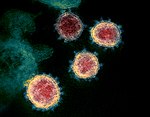 Severe acute respiratory syndrome coronavirus 2 Severe acute respiratory syndrome coronavirus 2 | ||||||||||||
| Method | single particle reconstruction / cryo EM / Resolution: 3.14 Å | ||||||||||||
 Authors Authors | Zhang J / Cai YF / Xiao TS / Rawson S / Peng HQ / Sterling SM / Walsh Jr RM / Volloch SR / Chen B | ||||||||||||
| Funding support |  United States, 3 items United States, 3 items
| ||||||||||||
 Citation Citation | Journal: Science / Year: 2021 Title: Structural basis for enhanced infectivity and immune evasion of SARS-CoV-2 variants. Authors: Yongfei Cai / Jun Zhang / Tianshu Xiao / Christy L Lavine / Shaun Rawson / Hanqin Peng / Haisun Zhu / Krishna Anand / Pei Tong / Avneesh Gautam / Shen Lu / Sarah M Sterling / Richard M Walsh ...Authors: Yongfei Cai / Jun Zhang / Tianshu Xiao / Christy L Lavine / Shaun Rawson / Hanqin Peng / Haisun Zhu / Krishna Anand / Pei Tong / Avneesh Gautam / Shen Lu / Sarah M Sterling / Richard M Walsh / Sophia Rits-Volloch / Jianming Lu / Duane R Wesemann / Wei Yang / Michael S Seaman / Bing Chen / Abstract: Several fast-spreading variants of severe acute respiratory syndrome coronavirus 2 (SARS-CoV-2) have become the dominant circulating strains in the COVID-19 pandemic. We report here cryo-electron ...Several fast-spreading variants of severe acute respiratory syndrome coronavirus 2 (SARS-CoV-2) have become the dominant circulating strains in the COVID-19 pandemic. We report here cryo-electron microscopy structures of the full-length spike (S) trimers of the B.1.1.7 and B.1.351 variants, as well as their biochemical and antigenic properties. Amino acid substitutions in the B.1.1.7 protein increase both the accessibility of its receptor binding domain and the binding affinity for receptor angiotensin-converting enzyme 2 (ACE2). The enhanced receptor engagement may account for the increased transmissibility. The B.1.351 variant has evolved to reshape antigenic surfaces of the major neutralizing sites on the S protein, making it resistant to some potent neutralizing antibodies. These findings provide structural details on how SARS-CoV-2 has evolved to enhance viral fitness and immune evasion. | ||||||||||||
| History |
|
- Structure visualization
| Movie |
Movie viewer |
|---|---|
| Structure viewer | EM map: SurfViewMolmilJmol/JSmol |
| Supplemental images |

- Downloads & links
-EMDB archive
| Map data | emd_24123.map.gz | 368.9 MB | EMDB map data format | |
|---|---|---|---|---|
| Header (meta data) | emd-24123-v30.xmlemd-24123.xml | 17.1 KB 17.1 KB | Display Display | EMDB header |
| Images |  emd_24123.png emd_24123.png | 88 KB | ||
| Others | emd_24123_additional_1.map.gz | 351.4 MB | ||
| Archive directory |  http://ftp.pdbj.org/pub/emdb/structures/EMD-24123ftp://ftp.pdbj.org/pub/emdb/structures/EMD-24123 http://ftp.pdbj.org/pub/emdb/structures/EMD-24123ftp://ftp.pdbj.org/pub/emdb/structures/EMD-24123 | HTTPS FTP |
-Related structure data
| Related structure data |  7n1uMC  7n1qC  7n1tC  7n1vC  7n1wC  7n1xC  7n1yC M: atomic model generated by this map C: citing same article ( |
|---|---|
| Similar structure data |


-Links
| EMDB pages | EMDB (EBI/PDBe) / EMDataResource |
|---|---|
| Related items in Molecule of the Month |


-Map
| File | Download / File: emd_24123.map.gz / Format: CCP4 / Size: 421.9 MB / Type: IMAGE STORED AS FLOATING POINT NUMBER (4 BYTES) | ||||||||||||||||||||||||||||||||||||||||||||||||||||||||||||||||||||
|---|---|---|---|---|---|---|---|---|---|---|---|---|---|---|---|---|---|---|---|---|---|---|---|---|---|---|---|---|---|---|---|---|---|---|---|---|---|---|---|---|---|---|---|---|---|---|---|---|---|---|---|---|---|---|---|---|---|---|---|---|---|---|---|---|---|---|---|---|---|
| Annotation | overall map | ||||||||||||||||||||||||||||||||||||||||||||||||||||||||||||||||||||
| Voxel size | X=Y=Z: 0.82507 Å | ||||||||||||||||||||||||||||||||||||||||||||||||||||||||||||||||||||
| Density |
| ||||||||||||||||||||||||||||||||||||||||||||||||||||||||||||||||||||
| Symmetry | Space group: 1 | ||||||||||||||||||||||||||||||||||||||||||||||||||||||||||||||||||||
| Details | EMDB XML:
CCP4 map header:
| ||||||||||||||||||||||||||||||||||||||||||||||||||||||||||||||||||||
-Supplemental data
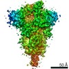
-Additional map: overall map with top region mask
| File | emd_24123_additional_1.map | ||||||||||||
|---|---|---|---|---|---|---|---|---|---|---|---|---|---|
| Annotation | overall map with top region mask | ||||||||||||
| Projections & Slices |
| ||||||||||||
| Density Histograms |
 Z
Z Y
Y X
X


- Sample components
-Entire : close state of pre-fusion SARS-CoV-2 B.1.1.7 spike variant glycop...
| Entire | Name: close state of pre-fusion SARS-CoV-2 B.1.1.7 spike variant glycoprotein |
|---|---|
| Components |
|
-Supramolecule #1: close state of pre-fusion SARS-CoV-2 B.1.1.7 spike variant glycop...
| Supramolecule | Name: close state of pre-fusion SARS-CoV-2 B.1.1.7 spike variant glycoprotein type: complex / ID: 1 / Parent: 0 / Macromolecule list: #1 Details: close state of pre-fusion SARS-CoV-2 B.1.1.7 spike variant glycoprotein |
|---|---|
| Source (natural) | Organism: Severe acute respiratory syndrome coronavirus 2 |
| Recombinant expression | Organism:  Homo sapiens (human) Homo sapiens (human) |
| Molecular weight | Theoretical: 440 kDa/nm |
-Macromolecule #1: Spike glycoprotein
| Macromolecule | Name: Spike glycoprotein / type: protein_or_peptide / ID: 1 / Number of copies: 3 / Enantiomer: LEVO |
|---|---|
| Source (natural) | Organism: Severe acute respiratory syndrome coronavirus 2 |
| Molecular weight | Theoretical: 144.3685 KDa |
| Recombinant expression | Organism: Homo sapiens (human) |
| Sequence | String: MFVFLVLLPL VSSQCVNLTT RTQLPPAYTN SFTRGVYYPD KVFRSSVLHS TQDLFLPFFS NVTWFHAISG TNGTKRFDNP VLPFNDGVY FASTEKSNII RGWIFGTTLD SKTQSLLIVN NATNVVIKVC EFQFCNDPFL GVYHKNNKSW MESEFRVYSS A NNCTFEYV ...String: MFVFLVLLPL VSSQCVNLTT RTQLPPAYTN SFTRGVYYPD KVFRSSVLHS TQDLFLPFFS NVTWFHAISG TNGTKRFDNP VLPFNDGVY FASTEKSNII RGWIFGTTLD SKTQSLLIVN NATNVVIKVC EFQFCNDPFL GVYHKNNKSW MESEFRVYSS A NNCTFEYV SQPFLMDLEG KQGNFKNLRE FVFKNIDGYF KIYSKHTPIN LVRDLPQGFS ALEPLVDLPI GINITRFQTL LA LHRSYLT PGDSSSGWTA GAAAYYVGYL QPRTFLLKYN ENGTITDAVD CALDPLSETK CTLKSFTVEK GIYQTSNFRV QPT ESIVRF PNITNLCPFG EVFNATRFAS VYAWNRKRIS NCVADYSVLY NSASFSTFKC YGVSPTKLND LCFTNVYADS FVIR GDEVR QIAPGQTGKI ADYNYKLPDD FTGCVIAWNS NNLDSKVGGN YNYLYRLFRK SNLKPFERDI STEIYQAGST PCNGV EGFN CYFPLQSYGF QPTYGVGYQP YRVVVLSFEL LHAPATVCGP KKSTNLVKNK CVNFNFNGLT GTGVLTESNK KFLPFQ QFG RDIDDTTDAV RDPQTLEILD ITPCSFGGVS VITPGTNTSN QVAVLYQGVN CTEVPVAIHA DQLTPTWRVY STGSNVF QT RAGCLIGAEH VNNSYECDIP IGAGICASYQ TQTNSHRRAR SVASQSIIAY TMSLGAENSV AYSNNSIAIP INFTISVT T EILPVSMTKT SVDCTMYICG DSTECSNLLL QYGSFCTQLN RALTGIAVEQ DKNTQEVFAQ VKQIYKTPPI KDFGGFNFS QILPDPSKPS KRSFIEDLLF NKVTLADAGF IKQYGDCLGD IAARDLICAQ KFNGLTVLPP LLTDEMIAQY TSALLAGTIT SGWTFGAGA ALQIPFAMQM AYRFNGIGVT QNVLYENQKL IANQFNSAIG KIQDSLSSTA SALGKLQDVV NQNAQALNTL V KQLSSNFG AISSVLNDIL ARLDKVEAEV QIDRLITGRL QSLQTYVTQQ LIRAAEIRAS ANLAATKMSE CVLGQSKRVD FC GKGYHLM SFPQSAPHGV VFLHVTYVPA QEKNFTTAPA ICHDGKAHFP REGVFVSNGT HWFVTQRNFY EPQIITTHNT FVS GNCDVV IGIVNNTVYD PLQPELDSFK EELDKYFKNH TSPDVDLGDI SGINASVVNI QKEIDRLNEV AKNLNESLID LQEL GKYEQ YIKWPWYIWL GFIAGLIAIV MVTIMLCCMT SCCSCLKGCC SCGSCCKFDE DDSEPVLKGV KLHYTSGGGS AWSHP QFEK GGGSGGGSGG SSAWSHPQFE K |
-Macromolecule #5: 2-acetamido-2-deoxy-beta-D-glucopyranose
| Macromolecule | Name: 2-acetamido-2-deoxy-beta-D-glucopyranose / type: ligand / ID: 5 / Number of copies: 24 / Formula: NAG |
|---|---|
| Molecular weight | Theoretical: 221.208 Da |
| Chemical component information |  ChemComp-NAG: |
-Experimental details
-Structure determination
| Method | cryo EM |
|---|---|
 Processing Processing | single particle reconstruction |
| Aggregation state | particle |
-Sample preparation
| Concentration | 1 mg/mL | ||||||||||||
|---|---|---|---|---|---|---|---|---|---|---|---|---|---|
| Buffer | pH: 7.5 Component:
| ||||||||||||
| Vitrification | Cryogen name: ETHANE / Chamber humidity: 100 % / Chamber temperature: 277.15 K / Instrument: FEI VITROBOT MARK IV |
- Electron microscopy
| Microscope | FEI TITAN KRIOS |
|---|---|
| Electron beam | Acceleration voltage: 300 kV / Electron source: FIELD EMISSION GUN |
| Electron optics | Illumination mode: FLOOD BEAM / Imaging mode: BRIGHT FIELDBright-field microscopy |
| Image recording | Film or detector model: GATAN K3 BIOQUANTUM (6k x 4k) / Average electron dose: 53.4 e/Å2 |
| Experimental equipment |  Model: Titan Krios / Image courtesy: FEI Company |
-Image processing
| Particle selection | Number selected: 2325106 |
|---|---|
| Startup model | Type of model: PDB ENTRY PDB model - PDB ID: |
| Initial angle assignment | Type: MAXIMUM LIKELIHOOD |
| Final 3D classification | Software - Name: RELION |
| Final angle assignment | Type: MAXIMUM LIKELIHOOD |
| Final reconstruction | Resolution.type: BY AUTHOR / Resolution: 3.14 Å / Resolution method: FSC 0.143 CUT-OFF / Software - Name: RELION / Number images used: 41138 |

