 Movie
Movie Controller


+ Open data

- Basic information
| Entry | Database: EMDB / ID: EMD-23049 | |||||||||
|---|---|---|---|---|---|---|---|---|---|---|
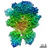
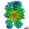
| Title | Cryo-EM structure of human Factor V at 3.6 Angstrom resolution | |||||||||
 Map data Map data | ||||||||||
 Sample Sample |
| |||||||||
| Function / homology |  Function and homology information Function and homology informationresponse to vitamin K /  platelet alpha granule / Cargo concentration in the ER / blood circulation / COPII-mediated vesicle transport / COPII-coated ER to Golgi transport vesicle / Common Pathway of Fibrin Clot Formation / endoplasmic reticulum-Golgi intermediate compartment membrane / platelet alpha granule lumen / Post-translational protein phosphorylation ...response to vitamin K / platelet alpha granule / Cargo concentration in the ER / blood circulation / COPII-mediated vesicle transport / COPII-coated ER to Golgi transport vesicle / Common Pathway of Fibrin Clot Formation / endoplasmic reticulum-Golgi intermediate compartment membrane / platelet alpha granule lumen / Post-translational protein phosphorylation / Regulation of Insulin-like Growth Factor (IGF) transport and uptake by Insulin-like Growth Factor Binding Proteins (IGFBPs) / extracellular vesicle / blood coagulation / Platelet degranulation / copper ion binding / endoplasmic reticulum lumen / extracellular space / extracellular region / membrane / plasma membrane platelet alpha granule / Cargo concentration in the ER / blood circulation / COPII-mediated vesicle transport / COPII-coated ER to Golgi transport vesicle / Common Pathway of Fibrin Clot Formation / endoplasmic reticulum-Golgi intermediate compartment membrane / platelet alpha granule lumen / Post-translational protein phosphorylation ...response to vitamin K / platelet alpha granule / Cargo concentration in the ER / blood circulation / COPII-mediated vesicle transport / COPII-coated ER to Golgi transport vesicle / Common Pathway of Fibrin Clot Formation / endoplasmic reticulum-Golgi intermediate compartment membrane / platelet alpha granule lumen / Post-translational protein phosphorylation / Regulation of Insulin-like Growth Factor (IGF) transport and uptake by Insulin-like Growth Factor Binding Proteins (IGFBPs) / extracellular vesicle / blood coagulation / Platelet degranulation / copper ion binding / endoplasmic reticulum lumen / extracellular space / extracellular region / membrane / plasma membraneSimilarity search - Function | |||||||||
| Biological species |  Homo sapiens (human) / Human (human) Homo sapiens (human) / Human (human) | |||||||||
| Method | single particle reconstruction / cryo EM / Resolution: 3.6 Å | |||||||||
 Authors Authors | Ruben EA / Di Cera E | |||||||||
| Funding support |  United States, 1 items United States, 1 items
| |||||||||
 Citation Citation | Journal: Blood / Year: 2021 Title: Cryo-EM structures of human coagulation factors V and Va. Authors: Eliza A Ruben / Michael J Rau / James A J Fitzpatrick / Enrico Di Cera / Abstract: Coagulation factor V (fV) is the precursor of fVa, which, together with fXa, Ca2+, and phospholipids, defines the prothrombinase complex and activates prothrombin in the penultimate step of the ...Coagulation factor V (fV) is the precursor of fVa, which, together with fXa, Ca2+, and phospholipids, defines the prothrombinase complex and activates prothrombin in the penultimate step of the coagulation cascade. We solved the cryogenic electron microscopy (cryo-EM) structures of human fV and fVa at atomic (3.3 Å) and near-atomic (4.4 Å) resolution, respectively. The structure of fV reveals the entire A1-A2-B-A3-C1-C2 assembly, but with a surprisingly disordered B domain. The C1 and C2 domains provide a platform for interaction with phospholipid membranes and support the A1 and A3 domains, with the A2 domain sitting on top of them. The B domain is highly dynamic and visible only for short segments connecting to the A2 and A3 domains. The A2 domain reveals all sites of proteolytic processing by thrombin and activated protein C, a partially buried epitope for binding fXa, and fully exposed epitopes for binding activated protein C and prothrombin. Removal of the B domain and activation to fVa exposes the sites of cleavage by activated protein C at R306 and R506 and produces increased disorder in the A1-A2-A3-C1-C2 assembly, especially in the C-terminal acidic portion of the A2 domain that is responsible for prothrombin binding. Ordering of this region and full exposure of the fXa epitope emerge as necessary steps in the assembly of the prothrombin-prothrombinase complex. These structures offer molecular context for the function of fV and fVa and pioneer the analysis of coagulation factors by cryo-EM. | |||||||||
| History |
|
- Structure visualization
| Movie |
Movie viewer |
|---|---|
| Structure viewer | EM map: SurfViewMolmilJmol/JSmol |
| Supplemental images |

- Downloads & links
-EMDB archive
| Map data | emd_23049.map.gz | 93.5 MB | EMDB map data format | |
|---|---|---|---|---|
| Header (meta data) | emd-23049-v30.xmlemd-23049.xml | 14.7 KB 14.7 KB | Display Display | EMDB header |
| Images | 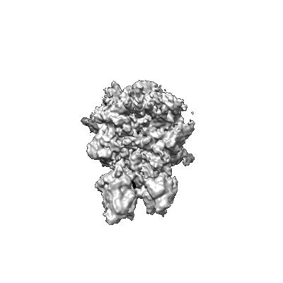 emd_23049.png emd_23049.png | 44.5 KB | ||
| Masks | emd_23049_msk_1.map | 98.9 MB | Mask map | |
| Others | emd_23049_half_map_1.map.gzemd_23049_half_map_2.map.gz | 91.7 MB 91.7 MB | ||
| Archive directory |  http://ftp.pdbj.org/pub/emdb/structures/EMD-23049ftp://ftp.pdbj.org/pub/emdb/structures/EMD-23049 http://ftp.pdbj.org/pub/emdb/structures/EMD-23049ftp://ftp.pdbj.org/pub/emdb/structures/EMD-23049 | HTTPS FTP |
-Related structure data
| Related structure data |  7kvfMC  7kveC  7kxyC C: citing same article ( M: atomic model generated by this map |
|---|---|
| Similar structure data |


-Links
| EMDB pages | EMDB (EBI/PDBe) / EMDataResource |
|---|---|
| Related items in Molecule of the Month |


-Map
| File | Download / File: emd_23049.map.gz / Format: CCP4 / Size: 98.9 MB / Type: IMAGE STORED AS FLOATING POINT NUMBER (4 BYTES) | ||||||||||||||||||||||||||||||||||||||||||||||||||||||||||||||||||||
|---|---|---|---|---|---|---|---|---|---|---|---|---|---|---|---|---|---|---|---|---|---|---|---|---|---|---|---|---|---|---|---|---|---|---|---|---|---|---|---|---|---|---|---|---|---|---|---|---|---|---|---|---|---|---|---|---|---|---|---|---|---|---|---|---|---|---|---|---|---|
| Voxel size | X=Y=Z: 1.1 Å | ||||||||||||||||||||||||||||||||||||||||||||||||||||||||||||||||||||
| Density |
| ||||||||||||||||||||||||||||||||||||||||||||||||||||||||||||||||||||
| Symmetry | Space group: 1 | ||||||||||||||||||||||||||||||||||||||||||||||||||||||||||||||||||||
| Details | EMDB XML:
CCP4 map header:
| ||||||||||||||||||||||||||||||||||||||||||||||||||||||||||||||||||||
-Supplemental data
-Mask #1
| File | emd_23049_msk_1.map | ||||||||||||
|---|---|---|---|---|---|---|---|---|---|---|---|---|---|
| Projections & Slices |
| ||||||||||||
| Density Histograms |
 Z
Z Y
Y X
X


-Half map: #2
| File | emd_23049_half_map_1.map | ||||||||||||
|---|---|---|---|---|---|---|---|---|---|---|---|---|---|
| Projections & Slices |
| ||||||||||||
| Density Histograms |


-Half map: #1
| File | emd_23049_half_map_2.map | ||||||||||||
|---|---|---|---|---|---|---|---|---|---|---|---|---|---|
| Projections & Slices |
| ||||||||||||
| Density Histograms |


- Sample components
-Entire : Human Factor V
| Entire | Name: Human Factor V |
|---|---|
| Components |
|
-Supramolecule #1: Human Factor V
| Supramolecule | Name: Human Factor V / type: complex / ID: 1 / Parent: 0 / Macromolecule list: all |
|---|---|
| Source (natural) | Organism: Homo sapiens (human) |
-Macromolecule #1: Coagulation factor V
| Macromolecule | Name: Coagulation factor V / type: protein_or_peptide / ID: 1 / Number of copies: 1 / Enantiomer: LEVO |
|---|---|
| Source (natural) | Organism: Human (human) |
| Molecular weight | Theoretical: 248.973234 KDa |
| Sequence | String: AQLRQFYVAA QGISWSYRPE PTNSSLNLSV TSFKKIVYRE YEPYFKKEKP QSTISGLLGP TLYAEVGDII KVHFKNKADK PLSIHPQGI RYSKLSEGAS YLDHTFPAEK MDDAVAPGRE YTYEWSISED SGPTHDDPPC LTHIYYSHEN LIEDFNSGLI G PLLICKKG ...String: AQLRQFYVAA QGISWSYRPE PTNSSLNLSV TSFKKIVYRE YEPYFKKEKP QSTISGLLGP TLYAEVGDII KVHFKNKADK PLSIHPQGI RYSKLSEGAS YLDHTFPAEK MDDAVAPGRE YTYEWSISED SGPTHDDPPC LTHIYYSHEN LIEDFNSGLI G PLLICKKG TLTEGGTQKT FDKQIVLLFA VFDESKSWSQ SSSLMYTVNG YVNGTMPDIT VCAHDHISWH LLGMSSGPEL FS IHFNGQV LEQNHHKVSA ITLVSATSTT ANMTVGPEGK WIISSLTPKH LQAGMQAYID IKNCPKKTRN LKKITREQRR HMK RWEYFI AAEEVIWDYA PVIPANMDKK YRSQHLDNFS NQIGKHYKKV MYTQYEDESF TKHTVNPNMK EDGILGPIIR AQVR DTLKI VFKNMASRPY SIYPHGVTFS PYEDEVNSSF TSGRNNTMIR AVQPGETYTY KWNILEFDEP TENDAQCLTR PYYSD VDIM RDIASGLIGL LLICKSRSLD RRGIQRAADI EQQAVFAVFD ENKSWYLEDN INKFCENPDE VKRDDPKFYE SNIMST ING YVPESITTLG FCFDDTVQWH FCSVGTQNEI LTIHFTGHSF IYGKRHEDTL TLFPMRGESV TVTMDNVGTW MLTSMNS SP RSKKLRLKFR DVKCIPDDDE DSYEIFEPPE STVMATRKMH DRLEPEDEES DADYDYQNRL AAALGIRSFR NSSLNQEE E EFNLTALALE NGTEFVSSNT DIIVGSNYSS PSNISKFTVN NLAEPQKAPS HQQATTAGSP LRHLIGKNSV LNSSTAEHS SPYSEDPIED PLQPDVTGIR LLSLGAGEFK SQEHAKHKGP KVERDQAAKH RFSWMKLLAH KVGRHLSQDT GSPSGMRPWE DLPSQDTGS PSRMRPWKDP PSDLLLLKQS NSSKILVGRW HLASEKGSYE IIQDTDEDTA VNNWLISPQN ASRAWGESTP L ANKPGKQS GHPKFPRVRH KSLQVRQDGG KSRLKKSQFL IKTRKKKKEK HTHHAPLSPR TFHPLRSEAY NTFSERRLKH SL VLHKSNE TSLPTDLNQT LPSMDFGWIA SLPDHNQNSS NDTGQASCPP GLYQTVPPEE HYQTFPIQDP DQMHSTSDPS HRS SSPELS EMLEYDRSHK SFPTDISQMS PSSEHEVWQT VISPDLSQVT LSPELSQTNL SPDLSHTTLS PELIQRNLSP ALGQ MPISP DLSHTTLSPD LSHTTLSLDL SQTNLSPELS QTNLSPALGQ MPLSPDLSHT TLSLDFSQTN LSPELSHMTL SPELS QTNL SPALGQMPIS PDLSHTTLSL DFSQTNLSPE LSQTNLSPAL GQMPLSPDPS HTTLSLDLSQ TNLSPELSQT NLSPDL SEM PLFADLSQIP LTPDLDQMTL SPDLGETDLS PNFGQMSLSP DLSQVTLSPD ISDTTLLPDL SQISPPPDLD QIFYPSE SS QSLLLQEFNE SFPYPDLGQM PSPSSPTLND TFLSKEFNPL VIVGLSKDGT DYIEIIPKEE VQSSEDDYAE IDYVPYDD P YKTDVRTNIN SSRDPDNIAA WYLRSNNGNR RNYYIAAEEI SWDYSEFVQR ETDIEDSDDI PEDTTYKKVV FRKYLDSTF TKRDPRGEYE EHLGILGPII RAEVDDVIQV RFKNLASRPY SLHAHGLSYE KSSEGKTYED DSPEWFKEDN AVQPNSSYTY VWHATERSG PESPGSACRA WAYYSAVNPE KDIHSGLIGP LLICQKGILH KDSNMPMDMR EFVLLFMTFD EKKSWYYEKK S RSSWRLTS SEMKKSHEFH AINGMIYSLP GLKMYEQEWV RLHLLNIGGS QDIHVVHFHG QTLLENGNKQ HQLGVWPLLP GS FKTLEMK ASKPGWWLLN TEVGENQRAG MQTPFLIMDR DCRMPMGLST GIISDSQIKA SEFLGYWEPR LARLNNGGSY NAW SVEKLA AEFASKPWIQ VDMQKEVIIT GIQTQGAKHY LKSCYTTEFY VAYSSNQINW QIFKGNSTRN VMYFNGNSDA STIK ENQFD PPIVARYIRI SPTRAYNRPT LRLELQGCEV NGCSTPLGME NGKIENKQIT ASSFKKSWWG DYWEPFRARL NAQGR VNAW QAKANNNKQW LEIDLLKIKK ITAIITQGCK SLSSEMYVKS YTIHYSEQGV EWKPYRLKSS MVDKIFEGNT NTKGHV KNF FNPPIISRFI RVIPKTWNQS IALRLELFGC DIY |
-Experimental details
-Structure determination
| Method | cryo EM |
|---|---|
 Processing Processing | single particle reconstruction |
| Aggregation state | particle |
-Sample preparation
| Buffer | pH: 7.4 / Details: 20 mM HEPES, 150 mM NaCl, 5 mM CaCl2 |
|---|---|
| Vitrification | Cryogen name: ETHANE |
- Electron microscopy
| Microscope | FEI TITAN KRIOS |
|---|---|
| Electron beam | Acceleration voltage: 300 kV / Electron source: FIELD EMISSION GUN |
| Electron optics | Illumination mode: OTHER / Imaging mode: BRIGHT FIELDBright-field microscopy |
| Image recording | Film or detector model: GATAN K2 QUANTUM (4k x 4k) / Average electron dose: 1.65 e/Å2 |
| Experimental equipment |  Model: Titan Krios / Image courtesy: FEI Company |
-Image processing
| Initial angle assignment | Type: ANGULAR RECONSTITUTION |
|---|---|
| Final angle assignment | Type: ANGULAR RECONSTITUTION |
| Final reconstruction | Resolution.type: BY AUTHOR / Resolution: 3.6 Å / Resolution method: FSC 0.143 CUT-OFF / Number images used: 299182 |
