 Movie
Movie Controller

[English] 日本語
 Yorodumi
- EMDB-21310: Cryo-EM map of C-terminal half of Leucine Rich Repeat Kinase 2 as... -
+ Open data

- Basic information
| Entry | Database: EMDB / ID: EMD-21310 | ||||||||||||
|---|---|---|---|---|---|---|---|---|---|---|---|---|---|
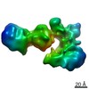
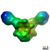
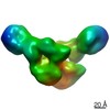
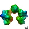
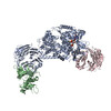
| Title | Cryo-EM map of C-terminal half of Leucine Rich Repeat Kinase 2 as a WD40-WD40 domain dimer at 13.4 angstroms | ||||||||||||
 Map data Map data | 13.39A map of Leucine Rich Repeat Kinase 2 truncate as a WD40 mediated dimer | ||||||||||||
 Sample Sample |
| ||||||||||||
| Biological species |   Homo sapiens (human) Homo sapiens (human) | ||||||||||||
| Method | single particle reconstruction / cryo EM / Resolution: 13.4 Å | ||||||||||||
 Authors Authors | Leschziner A / Deniston C / Lahiri I | ||||||||||||
| Funding support |  United States, 3 items United States, 3 items
| ||||||||||||
 Citation Citation | Journal: Nature / Year: 2020 Title: Structure of LRRK2 in Parkinson's disease and model for microtubule interaction. Authors: C K Deniston / J Salogiannis / S Mathea / D M Snead / I Lahiri / M Matyszewski / O Donosa / R Watanabe / J Böhning / A K Shiau / S Knapp / E Villa / S L Reck-Peterson / A E Leschziner /    Abstract: Leucine-rich repeat kinase 2 (LRRK2) is the most commonly mutated gene in familial Parkinson's disease and is also linked to its idiopathic form. LRRK2 has been proposed to function in membrane ...Leucine-rich repeat kinase 2 (LRRK2) is the most commonly mutated gene in familial Parkinson's disease and is also linked to its idiopathic form. LRRK2 has been proposed to function in membrane trafficking and colocalizes with microtubules. Despite the fundamental importance of LRRK2 for understanding and treating Parkinson's disease, structural information on the enzyme is limited. Here we report the structure of the catalytic half of LRRK2, and an atomic model of microtubule-associated LRRK2 built using a reported cryo-electron tomography in situ structure. We propose that the conformation of the LRRK2 kinase domain regulates its interactions with microtubules, with a closed conformation favouring oligomerization on microtubules. We show that the catalytic half of LRRK2 is sufficient for filament formation and blocks the motility of the microtubule-based motors kinesin 1 and cytoplasmic dynein 1 in vitro. Kinase inhibitors that stabilize an open conformation relieve this interference and reduce the formation of LRRK2 filaments in cells, whereas inhibitors that stabilize a closed conformation do not. Our findings suggest that LRRK2 can act as a roadblock for microtubule-based motors and have implications for the design of therapeutic LRRK2 kinase inhibitors. | ||||||||||||
| History |
|
- Structure visualization
| Movie |
 Movie viewer Movie viewer |
|---|---|
| Structure viewer | EM map: SurfViewMolmilJmol/JSmol |
| Supplemental images |

- Downloads & links
-EMDB archive
| Map data | emd_21310.map.gz | 9.4 MB | EMDB map data format | |
|---|---|---|---|---|
| Header (meta data) | emd-21310-v30.xmlemd-21310.xml | 20 KB 20 KB | Display Display | EMDB header |
| FSC (resolution estimation) | emd_21310_fsc.xml | 6.8 KB | Display | FSC data file |
| Images |  emd_21310.png emd_21310.png | 81.4 KB | ||
| Others | emd_21310_half_map_1.map.gzemd_21310_half_map_2.map.gz | 17.9 MB 17.9 MB | ||
| Archive directory |  http://ftp.pdbj.org/pub/emdb/structures/EMD-21310ftp://ftp.pdbj.org/pub/emdb/structures/EMD-21310 http://ftp.pdbj.org/pub/emdb/structures/EMD-21310ftp://ftp.pdbj.org/pub/emdb/structures/EMD-21310 | HTTPS FTP |
-Related structure data


-Links
| EMDB pages | EMDB (EBI/PDBe) / EMDataResource |
|---|
-Map
| File | Download / File: emd_21310.map.gz / Format: CCP4 / Size: 19.4 MB / Type: IMAGE STORED AS FLOATING POINT NUMBER (4 BYTES) | ||||||||||||||||||||||||||||||||||||||||||||||||||||||||||||
|---|---|---|---|---|---|---|---|---|---|---|---|---|---|---|---|---|---|---|---|---|---|---|---|---|---|---|---|---|---|---|---|---|---|---|---|---|---|---|---|---|---|---|---|---|---|---|---|---|---|---|---|---|---|---|---|---|---|---|---|---|---|
| Annotation | 13.39A map of Leucine Rich Repeat Kinase 2 truncate as a WD40 mediated dimer | ||||||||||||||||||||||||||||||||||||||||||||||||||||||||||||
| Voxel size | X=Y=Z: 2.32 Å | ||||||||||||||||||||||||||||||||||||||||||||||||||||||||||||
| Density |
| ||||||||||||||||||||||||||||||||||||||||||||||||||||||||||||
| Symmetry | Space group: 1 | ||||||||||||||||||||||||||||||||||||||||||||||||||||||||||||
| Details | EMDB XML:
CCP4 map header:
| ||||||||||||||||||||||||||||||||||||||||||||||||||||||||||||
-Supplemental data
-Half map: Half map 2
| File | emd_21310_half_map_1.map | ||||||||||||
|---|---|---|---|---|---|---|---|---|---|---|---|---|---|
| Annotation | Half map 2 | ||||||||||||
| Projections & Slices |
| ||||||||||||
| Density Histograms |
 Z
Z Y
Y X
X


-Half map: Half map 1
| File | emd_21310_half_map_2.map | ||||||||||||
|---|---|---|---|---|---|---|---|---|---|---|---|---|---|
| Annotation | Half map 1 | ||||||||||||
| Projections & Slices |
| ||||||||||||
| Density Histograms |


- Sample components
-Entire : Truncated Leucine Rich Repeat Kinase 2 WD40-WD40 domain mediated ...
| Entire | Name: Truncated Leucine Rich Repeat Kinase 2 WD40-WD40 domain mediated dimer. |
|---|---|
| Components |
|
-Supramolecule #1: Truncated Leucine Rich Repeat Kinase 2 WD40-WD40 domain mediated ...
| Supramolecule | Name: Truncated Leucine Rich Repeat Kinase 2 WD40-WD40 domain mediated dimer. type: organelle_or_cellular_component / ID: 1 / Parent: 0 / Macromolecule list: all Details: A WD40-WD40 (13.4A) mediated dimer of truncated Leucine Rich Repeat Kinase 2 starting at residue 1327 all the way to the C-terminal. |
|---|---|
| Source (natural) | Organism: Homo sapiens (human) |
| Molecular weight | Theoretical: 274 KDa |
| Recombinant expression | Organism:   Spodoptera frugiperda (fall armyworm) Spodoptera frugiperda (fall armyworm) |
-Macromolecule #1: Leucine Rich Repeat Kinase 2
| Macromolecule | Name: Leucine Rich Repeat Kinase 2 / type: protein_or_peptide / ID: 1 / Enantiomer: LEVO / EC number: non-specific serine/threonine protein kinase |
|---|---|
| Source (natural) | Organism: Homo sapiens (human) |
| Recombinant expression | Organism: Spodoptera frugiperda (fall armyworm) |
| Sequence | String: KKAVPYNRMK LMIVGN(TPO)GSG KTTLLQQLMK TKKSDLGMQS ATVGIDVKDW PIQIRDKRKR DLVLNVWDFA GREEFYSTHP HFMTQRALYL AVYDLSKGQA EVDAMKPWLF NIKARASSSP VILVGTHLDV SDEKQRKACM SKITKELLNK RGFPAIRDYH FVNATEESDA ...String: KKAVPYNRMK LMIVGN(TPO)GSG KTTLLQQLMK TKKSDLGMQS ATVGIDVKDW PIQIRDKRKR DLVLNVWDFA GREEFYSTHP HFMTQRALYL AVYDLSKGQA EVDAMKPWLF NIKARASSSP VILVGTHLDV SDEKQRKACM SKITKELLNK RGFPAIRDYH FVNATEESDA LAKLRKTIIN ESLNFKIRDQ LVVGQLIPDC YVELEKIILS ERKNVPIEFP VIDRKRLLQL VRENQLQLDE NELPHAVHFL NESGVLLHFQ DPALQLSDLY FVEPKWLCKI MAQILTVKVE GCPKHPKGII SRRDVEKFLS KKRKFPKNYM SQYFKLLEKF QIALPIGEEY LLVPSSLSDH RPVIELPHCE NSEIIIRLYE MPYFPMGFWS RLINRLLEIS PYMLSGRERA LRPNRMYWRQ GIYLNWSPEA YCLVGSEVLD NHPESFLKIT VPSCRKGCIL LGQVVDHIDS LMEEWFPGLL EIDICGEGET LLKKWALYSF NDGEEHQKIL LDDLMKKAEE GDLLVNPDQP RLTIPISQIA PDLILADLPR NIMLNNDELE FEQAPEFLLG DGSFGSVYRA AYEGEEVAVK IFNKHTSLRL LRQELVVLCH LHHPSLISLL AAGIRPRMLV MELASKGSLD RLLQQDKASL TRTLQHRIAL HVADGLRYLH SAMIIYRDLK PHNVLLFTLY PNAAIIAKIA DYGIAQYCCR MGIKTSEGTP GFRAPEVARG NVIYNQQADV YSFGLLLYDI LTTGGRIVEG LKFPNEFDEL EIQGKLPDPV KEYGCAPWPM VEKLIKQCLK ENPQERPTSA QVFDILNSAE LVCLTRRILL PKNVIVECMV ATHHNSRNAS IWLGCGHTDR GQLSFLDLNT EGYTSEEVAD SRILCLALVH LPVEKESWIV SGTQSGTLLV INTEDGKKRH TLEKMTDSVT CLYCNSFSKQ SKQKNFLLVG TADGKLAIFE DKTVKLKGAA PLKILNIGNV STPLMCLSES TNSTERNVMW GGCGTKIFSF SNDFTIQKLI ETRTSQLFSY AAFSDSNIIT VVVDTALYIA KQNSPVVEVW DKKTEKLCGL IDCVHFLREV MVKENKESKH KMSYSGRVKT LCLQKNTALW IGTGGGHILL LDLSTRRLIR VIYNFCNSVR VMMTAQLGSL KNVMLVLGYN RKNTEGTQKQ KEIQSCLTVW DINLPHEVQN LEKHIEVRKE LAEKMRRTSV E |
-Experimental details
-Structure determination
| Method | cryo EM |
|---|---|
 Processing Processing | single particle reconstruction |
| Aggregation state | particle |
-Sample preparation
| Concentration | 0.6 mg/mL | |||||||||||||||||||||
|---|---|---|---|---|---|---|---|---|---|---|---|---|---|---|---|---|---|---|---|---|---|---|
| Buffer | pH: 7.4 Component:
Details: Some samples had the addition of 0.05mM Digitonin or 0.03% octyl glucoside detergents in order to combat preferred orientation. | |||||||||||||||||||||
| Grid | Model: Quantifoil, UltrAuFoil, R1.2/1.3 / Material: GOLD / Pretreatment - Type: GLOW DISCHARGE | |||||||||||||||||||||
| Vitrification | Cryogen name: ETHANE / Chamber humidity: 100 % / Chamber temperature: 277.15 K / Instrument: FEI VITROBOT MARK II | |||||||||||||||||||||
| Details | This sample was imaged over a number of datasets. Specimen concentrations ranged from 0.6(4uM)-1.7(12uM) mg/mL, however the average was 0.6 and thus was entered as the reported specimen concentration. |
- Electron microscopy
| Microscope | FEI TALOS ARCTICA |
|---|---|
| Electron beam | Acceleration voltage: 200 kV / Electron source: FIELD EMISSION GUN |
| Electron optics | Illumination mode: FLOOD BEAM / Imaging mode: BRIGHT FIELDBright-field microscopy / Cs: 2.7 mm / Nominal defocus max: 2.0 µm / Nominal defocus min: 1.0 µm |
| Sample stage | Specimen holder model: FEI TITAN KRIOS AUTOGRID HOLDER / Cooling holder cryogen: NITROGEN |
| Details | Datasets collected at 36kx magnification (either counting mode or super resolution mode). One dataset collected at 30 degree tilt. |
| Image recording | Film or detector model: GATAN K2 SUMMIT (4k x 4k) / Number real images: 3100 / Average electron dose: 4.6 e/Å2 Details: Datasets collected at 36kx magnification (either counting mode or super resolution mode). Only a single dose has been entered, however datasets were collected at various doses between 4.6 ...Details: Datasets collected at 36kx magnification (either counting mode or super resolution mode). Only a single dose has been entered, however datasets were collected at various doses between 4.6 and 7.8 electrons per angstrom squared. See method section of paper for more details. |
| Experimental equipment | 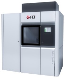 Model: Talos Arctica / Image courtesy: FEI Company |
-Image processing
| Particle selection | Number selected: 431387 Details: Cryolo Ver.1 {Wagner,2019} was used to pick particles. |
|---|---|
| CTF correction | Software - Name: CTFFIND (ver. 4) |
| Startup model | Type of model: OTHER Details: Initial model was built from our reconstruction of Leucine Rich Repeat Kinase 2 truncate fit into a ab initio map of the dimer out of Cryosparc2. See the paper for more details. |
| Initial angle assignment | Type: MAXIMUM LIKELIHOOD / Software - Name: cryoSPARC (ver. 2) |
| Final angle assignment | Type: MAXIMUM LIKELIHOOD / Software - Name: cryoSPARC (ver. 2) |
| Final reconstruction | Applied symmetry - Point group: C2 (2 fold cyclic) / Resolution.type: BY AUTHOR / Resolution: 13.4 Å / Resolution method: FSC 0.143 CUT-OFF / Software - Name: cryoSPARC (ver. 2) / Number images used: 63435 |
| FSC plot (resolution estimation) |  |
