 Movie
Movie Controller


+ Open data

- Basic information
| Entry | 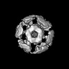 | |||||||||
|---|---|---|---|---|---|---|---|---|---|---|
| Title | nicotinic acetylcholine receptor in intact synaptic membrane | |||||||||
 Map data Map data | Map of acetylcholine receptor in membrane obtained from aligned volumes in helical reconstructions of tubular vesicles having different diameters and symmetry (see e.g. EMD-14942) | |||||||||
 Sample Sample |
| |||||||||
 Keywords Keywords | Ion channel / MEMBRANE PROTEIN | |||||||||
| Function / homology |  Function and homology information Function and homology informationacetylcholine-gated monoatomic cation-selective channel activity / transmembrane signaling receptor activity / postsynaptic membraneSimilarity search - Function | |||||||||
| Biological species |  Torpedo marmorata (marbled electric ray) / Torpedo marmorata (marbled electric ray) /  Tetronarce californica (Pacific electric ray) Tetronarce californica (Pacific electric ray) | |||||||||
| Method | single particle reconstruction / cryo EM / Resolution: 4.7 Å | |||||||||
 Authors Authors | Unwin N | |||||||||
| Funding support |  United Kingdom, 1 items United Kingdom, 1 items
| |||||||||
 Citation Citation | Journal: Proc Natl Acad Sci U S A / Year: 2024 Title: Influence of lipid bilayer on the structure of the muscle-type nicotinic acetylcholine receptor. Authors: Nigel Unwin / Abstract: The muscle-type nicotinic acetylcholine receptor is a transmitter-gated ion channel residing in the plasma membrane of electrocytes and striated muscle cells. It is present predominantly at synaptic ...The muscle-type nicotinic acetylcholine receptor is a transmitter-gated ion channel residing in the plasma membrane of electrocytes and striated muscle cells. It is present predominantly at synaptic junctions, where it effects rapid depolarization of the postsynaptic membrane in response to acetylcholine released into the synaptic cleft. Previously, cryo-EM of intact membrane from revealed that the lipid bilayer surrounding the junctional receptor has a uniquely asymmetric and ordered structure, due to a high concentration of cholesterol. It is now shown that this special lipid environment influences the transmembrane (TM) folding of the protein. All five submembrane MX helices of the membrane-intact junctional receptor align parallel to the surface of the cholesterol-ordered lipids in the inner leaflet of the bilayer; also, the TM helices in the outer leaflet are splayed apart. However in the structure obtained from the same protein after extraction and incorporation in nanodiscs, the MX helices do not align to a planar surface, and the TM helices arrange compactly in the outer leaflet. Realignment of the MX helices of the nanodisc-solved structure to a planar surface converts their adjoining TM helices into an obligatory splayed configuration, characteristic of the junctional receptor. Thus, the form of the receptor sustained by the special lipid environment of the synaptic junction is the one that mediates fast synaptic transmission; whereas, the nanodisc-embedded protein may be like the extrajunctional form, existing in a disordered lipid environment. | |||||||||
| History |
|
- Structure visualization
| Supplemental images |
|---|

- Downloads & links
-EMDB archive
| Map data | emd_18596.map.gz | 3.8 MB | EMDB map data format | |
|---|---|---|---|---|
| Header (meta data) | emd-18596-v30.xmlemd-18596.xml | 36.6 KB 36.6 KB | Display Display | EMDB header |
| Images |  emd_18596.png emd_18596.png | 51.5 KB | ||
| Filedesc metadata | emd-18596.cif.gz | 7.9 KB | ||
| Others | emd_18596_half_map_1.map.gzemd_18596_half_map_2.map.gz | 3.8 MB 3.8 MB | ||
| Archive directory |  http://ftp.pdbj.org/pub/emdb/structures/EMD-18596ftp://ftp.pdbj.org/pub/emdb/structures/EMD-18596 http://ftp.pdbj.org/pub/emdb/structures/EMD-18596ftp://ftp.pdbj.org/pub/emdb/structures/EMD-18596 | HTTPS FTP |
-Related structure data
| Related structure data | 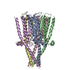 8qqmMC C: citing same article ( M: atomic model generated by this map |
|---|---|
| Similar structure data |


-Links
| EMDB pages | EMDB (EBI/PDBe) / EMDataResource |
|---|---|
| Related items in Molecule of the Month |


-Map
| File | Download / File: emd_18596.map.gz / Format: CCP4 / Size: 30.5 MB / Type: IMAGE STORED AS FLOATING POINT NUMBER (4 BYTES) | ||||||||||||||||||||||||||||||||||||
|---|---|---|---|---|---|---|---|---|---|---|---|---|---|---|---|---|---|---|---|---|---|---|---|---|---|---|---|---|---|---|---|---|---|---|---|---|---|
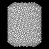
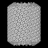
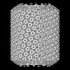
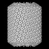
| Annotation | Map of acetylcholine receptor in membrane obtained from aligned volumes in helical reconstructions of tubular vesicles having different diameters and symmetry (see e.g. EMD-14942) | ||||||||||||||||||||||||||||||||||||
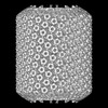
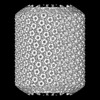
| Projections & slices | Image control
Images are generated by Spider. | ||||||||||||||||||||||||||||||||||||
| Voxel size | X=Y=Z: 1.34 Å | ||||||||||||||||||||||||||||||||||||
| Density |
| ||||||||||||||||||||||||||||||||||||
| Symmetry | Space group: 1 | ||||||||||||||||||||||||||||||||||||
| Details | EMDB XML:
|
 Z (Sec.)
 Y (Row.)
 X (Col.)


-Supplemental data
-Half map: First half map
| File | emd_18596_half_map_1.map | ||||||||||||
|---|---|---|---|---|---|---|---|---|---|---|---|---|---|
| Annotation | First half map | ||||||||||||
| Projections & Slices |
| ||||||||||||
| Density Histograms |


-Half map: Second half map
| File | emd_18596_half_map_2.map | ||||||||||||
|---|---|---|---|---|---|---|---|---|---|---|---|---|---|
| Annotation | Second half map | ||||||||||||
| Projections & Slices |
| ||||||||||||
| Density Histograms |


- Sample components
-Entire : Muscle-type nicotinic acetylcholine receptor in intact synaptic m...
| Entire | Name: Muscle-type nicotinic acetylcholine receptor in intact synaptic membrane |
|---|---|
| Components |
|
-Supramolecule #1: Muscle-type nicotinic acetylcholine receptor in intact synaptic m...
| Supramolecule | Name: Muscle-type nicotinic acetylcholine receptor in intact synaptic membrane type: organelle_or_cellular_component / ID: 1 / Parent: 0 / Macromolecule list: all Details: Postsynaptic membranes were isolated from fresh electric organ and incubated in low salt buffer to form ordered arrays of acetylcholine receptors in tubular vesicles |
|---|---|
| Source (natural) | Organism: Torpedo marmorata (marbled electric ray) |
| Molecular weight | Theoretical: 290 KDa |
-Macromolecule #1: Acetylcholine receptor subunit alpha
| Macromolecule | Name: Acetylcholine receptor subunit alpha / type: protein_or_peptide / ID: 1 Details: The extracellular domain is excluded from this analysis Number of copies: 2 / Enantiomer: LEVO |
|---|---|
| Source (natural) | Organism: Tetronarce californica (Pacific electric ray) |
| Molecular weight | Theoretical: 50.168164 KDa |
| Sequence | String: SEHETRLVAN LLENYNKVIR PVEHHTHFVD ITVGLQLIQL ISVDEVNQIV ETNVRLRQQW IDVRLRWNPA DYGGIKKIRL PSDDVWLPD LVLYNNADGD FAIVHMTKLL LDYTGKIMWT PPAIFKSYCE IIVTHFPFDQ QNCTMKLGIW TYDGTKVSIS P ESDRPDLS ...String: SEHETRLVAN LLENYNKVIR PVEHHTHFVD ITVGLQLIQL ISVDEVNQIV ETNVRLRQQW IDVRLRWNPA DYGGIKKIRL PSDDVWLPD LVLYNNADGD FAIVHMTKLL LDYTGKIMWT PPAIFKSYCE IIVTHFPFDQ QNCTMKLGIW TYDGTKVSIS P ESDRPDLS TFMESGEWVM KDYRGWKHWV YYTCCPDTPY LDITYHFIMQ RIPLYFVVNV IIPCLLFSFL TGLVFYLPTD SG EKMTLSI SVLLSLTVFL LVIVELIPST SSAVPLIGKY MLFTMIFVIS SIIITVVVIN THHRSPSTHT MPQWVRKIFI DTI PNVMFF STMKRASKEK QENKIFADDI DISDISGKQV TGEVIFQTPL IKNPDVKSAI EGVKYIAEHM KSDEESSNAA EEWK YVAMV IDHILLCVFM LICIIGTVSV FAGRLIELSQ EG UniProtKB: Acetylcholine receptor subunit alpha |
-Macromolecule #2: Acetylcholine receptor subunit delta
| Macromolecule | Name: Acetylcholine receptor subunit delta / type: protein_or_peptide / ID: 2 Details: The extracellular domain is excluded from this analysis Number of copies: 1 / Enantiomer: LEVO |
|---|---|
| Source (natural) | Organism: Tetronarce californica (Pacific electric ray) |
| Molecular weight | Theoretical: 57.625711 KDa |
| Sequence | String: VNEEERLIND LLIVNKYNKH VRPVKHNNEV VNIALSLTLS NLISLKETDE TLTSNVWMDH AWYDHRLTWN ASEYSDISIL RLPPELVWI PDIVLQNNND GQYHVAYFCN VLVRPNGYVT WLPPAIFRSS CPINVLYFPF DWQNCSLKFT ALNYDANEIT M DLMTDTID ...String: VNEEERLIND LLIVNKYNKH VRPVKHNNEV VNIALSLTLS NLISLKETDE TLTSNVWMDH AWYDHRLTWN ASEYSDISIL RLPPELVWI PDIVLQNNND GQYHVAYFCN VLVRPNGYVT WLPPAIFRSS CPINVLYFPF DWQNCSLKFT ALNYDANEIT M DLMTDTID GKDYPIEWII IDPEAFTENG EWEIIHKPAK KNIYPDKFPN GTNYQDVTFY LIIRRKPLFY VINFITPCVL IS FLASLAF YLPAESGEKM STAISVLLAQ AVFLLLTSQR LPETALAVPL IGKYLMFIMS LVTGVIVNCG IVLNFHFRTP STH VLSTRV KQIFLEKLPR ILHMSRADES EQPDWQNDLK LRRSSSVGYI SKAQEYFNIK SRSELMFEKQ SERHGLVPRV TPRI GFGNN NENIAASDQL HDEIKSGIDS TNYIVKQIKE KNAYDEEVGN WNLVGQTIDR LSMFIITPVM VLGTIFIFVM GNFNH PPAK PFEGDPFDYS SDHPRCA UniProtKB: Acetylcholine receptor subunit delta |
-Macromolecule #3: Acetylcholine receptor subunit beta
| Macromolecule | Name: Acetylcholine receptor subunit beta / type: protein_or_peptide / ID: 3 Details: The extracellular domain is excluded from this analysis Number of copies: 1 / Enantiomer: LEVO |
|---|---|
| Source (natural) | Organism: Tetronarce californica (Pacific electric ray) |
| Molecular weight | Theoretical: 53.731773 KDa |
| Sequence | String: SVMEDTLLSV LFETYNPKVR PAQTVGDKVT VRVGLTLTNL LILNEKIEEM TTNVFLNLAW TDYRLQWDPA AYEGIKDLRI PSSDVWQPD IVLMNNNDGS FEITLHVNVL VQHTGAVSWQ PSAIYRSSCT IKVMYFPFDW QNCTMVFKSY TYDTSEVTLQ H ALDAKGER ...String: SVMEDTLLSV LFETYNPKVR PAQTVGDKVT VRVGLTLTNL LILNEKIEEM TTNVFLNLAW TDYRLQWDPA AYEGIKDLRI PSSDVWQPD IVLMNNNDGS FEITLHVNVL VQHTGAVSWQ PSAIYRSSCT IKVMYFPFDW QNCTMVFKSY TYDTSEVTLQ H ALDAKGER EVKEIVINKD AFTENGQWSI EHKPSRKNWR SDDPSYEDVT FYLIIQRKPL FYIVYTIIPC ILISILAILV FY LPPDAGE KMSLSISALL AVTVFLLLLA DKVPETSLSV PIIIRYLMFI MILVAFSVIL SVVVLNLHHR SPNTHTMPNW IRQ IFIETL PPFLWIQRPV TTPSPDSKPT IISRANDEYF IRKPAGDFVC PVDNARVAVQ PERLFSEMKW HLNGLTQPVT LPQD LKEAV EAIKYIAEQL ESASEFDDLK KDWQYVAMVA DRLFLYVFFV ICSIGTFSIF LDASHNVPPD NPFA UniProtKB: Acetylcholine receptor subunit beta |
-Macromolecule #4: Acetylcholine receptor subunit gamma
| Macromolecule | Name: Acetylcholine receptor subunit gamma / type: protein_or_peptide / ID: 4 Details: The extracellular domain is excluded from this analysis Number of copies: 1 / Enantiomer: LEVO |
|---|---|
| Source (natural) | Organism: Tetronarce californica (Pacific electric ray) |
| Molecular weight | Theoretical: 56.335684 KDa |
| Sequence | String: ENEEGRLIEK LLGDYDKRII PAKTLDHIID VTLKLTLTNL ISLNEKEEAL TTNVWIEIQW NDYRLSWNTS EYEGIDLVRI PSELLWLPD VVLENNVDGQ FEVAYYANVL VYNDGSMYWL PPAIYRSTCP IAVTYFPFDW QNCSLVFRSQ TYNAHEVNLQ L SAEEGEAV ...String: ENEEGRLIEK LLGDYDKRII PAKTLDHIID VTLKLTLTNL ISLNEKEEAL TTNVWIEIQW NDYRLSWNTS EYEGIDLVRI PSELLWLPD VVLENNVDGQ FEVAYYANVL VYNDGSMYWL PPAIYRSTCP IAVTYFPFDW QNCSLVFRSQ TYNAHEVNLQ L SAEEGEAV EWIHIDPEDF TENGEWTIRH RPAKKNYNWQ LTKDDTDFQE IIFFLIIQRK PLFYIINIIA PCVLISSLVV LV YFLPAQA GGQKCTLSIS VLLAQTIFLF LIAQKVPETS LNVPLIGKYL IFVMFVSMLI VMNCVIVLNV SLRTPNTHSL SEK IKHLFL GFLPKYLGMQ LEPSEETPEK PQPRRRSSFG IMIKAEEYIL KKPRSELMFE EQKDRHGLKR VNKMTSDIDI GTTV DLYKD LANFAPEIKS CVEACNFIAK STKEQNDSGS ENENWVLIGK VIDKACFWIA LLLFSIGTLA IFLTGHFNQV PEFPF PGDP RKYVP UniProtKB: Acetylcholine receptor subunit gamma |
-Experimental details
-Structure determination
| Method | cryo EM |
|---|---|
 Processing Processing | single particle reconstruction |
| Aggregation state | helical array |
-Sample preparation
| Buffer | pH: 7 Component:
| |||||||||
|---|---|---|---|---|---|---|---|---|---|---|
| Grid | Model: Quantifoil R1.2/1.3 / Material: COPPER / Mesh: 300 / Support film - Material: CARBON / Support film - topology: HOLEY ARRAY / Support film - Film thickness: 20 / Pretreatment - Type: GLOW DISCHARGE / Pretreatment - Time: 60 sec. / Pretreatment - Atmosphere: AMYLAMINE Details: Grids were washed extensively in chloroform and coated with additional carbon before use | |||||||||
| Vitrification | Cryogen name: ETHANE / Chamber humidity: 95 % / Chamber temperature: 283 K / Instrument: HOMEMADE PLUNGER | |||||||||
| Details | Specimen comprises tubular vesicles which are imaged in thin ice over holes in the support film |
- Electron microscopy
| Microscope | FEI TITAN KRIOS |
|---|---|
| Electron beam | Acceleration voltage: 300 kV / Electron source: FIELD EMISSION GUN |
| Electron optics | C2 aperture diameter: 50.0 µm / Calibrated defocus max: 2.8000000000000003 µm / Calibrated defocus min: 1.2 µm / Calibrated magnification: 104478 / Illumination mode: FLOOD BEAM / Imaging mode: BRIGHT FIELDBright-field microscopy / Cs: 2.7 mm / Nominal defocus max: 2.8000000000000003 µm / Nominal defocus min: 1.2 µm / Nominal magnification: 59000 |
| Sample stage | Specimen holder model: FEI TITAN KRIOS AUTOGRID HOLDER / Cooling holder cryogen: NITROGEN |
| Temperature | Min: 70.0 K / Max: 70.0 K |
| Details | All images taken manually: by searching at low magnification for long straight and narrow tubes, then recording in integrating mode |
| Image recording | Film or detector model: FEI FALCON III (4k x 4k) / Detector mode: INTEGRATING / Number grids imaged: 200 / Number real images: 4045 / Average exposure time: 2.0 sec. / Average electron dose: 40.0 e/Å2 Details: Images were collected in integrating mode, 2 seconds exposure |
| Experimental equipment |  Model: Titan Krios / Image courtesy: FEI Company |
-Image processing
| Particle selection | Number selected: 160652 Details: 160652 tube segments from 4045 selected micrographs were reduced to 107524 tube segments after 2D classification |
|---|---|
| Startup model | Type of model: OTHER Details: Original model was a map of the tube obtained by Fourier-Bessel reconstruction |
| Initial angle assignment | Type: NOT APPLICABLE |
| Final 3D classification | Number classes: 34 / Avg.num./class: 3162 / Software - Name: RELION (ver. 2.1) |
| Final angle assignment | Type: NOT APPLICABLE |
| Final reconstruction | Number classes used: 34 / Applied symmetry - Point group: C1 (asymmetric) / Algorithm: FOURIER SPACE / Resolution.type: BY AUTHOR / Resolution: 4.7 Å / Resolution method: FSC 0.143 CUT-OFF / Software - Name: RELION (ver. 2.1) Details: The volumes for FSC determination were cut out from helical reconstructions of each class average Number images used: 107524 |
| Details | Images of appropriate helical families were selected on the basis of their FFTs, then drift-corrected and dose-weighted |
-Atomic model buiding 1
| Initial model |
| ||||||||||||||||||
|---|---|---|---|---|---|---|---|---|---|---|---|---|---|---|---|---|---|---|---|
| Details | Refinement parameters were chosen to minimise changes to the original secondary structure | ||||||||||||||||||
| Refinement | Space: REAL / Protocol: FLEXIBLE FIT / Overall B value: 350 | ||||||||||||||||||
| Output model | PDB-8qqm: |

