 Movie
Movie Controller

[English] 日本語
 Yorodumi
- EMDB-18493: Cryo-electron tomogram of lift-out lamella from cell-derived extr... -
+ Open data

- Basic information
| Entry | 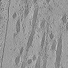 | |||||||||||||||
|---|---|---|---|---|---|---|---|---|---|---|---|---|---|---|---|---|
| Title | Cryo-electron tomogram of lift-out lamella from cell-derived extracellular matrix (example 4) | |||||||||||||||
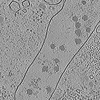
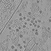
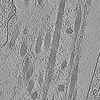
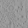
 Map data Map data | Tomogram acquired on cryo-lift-out lamella of a cell-derived matrix showing mainly extracellular structures such as collagen. | |||||||||||||||
 Sample Sample |
| |||||||||||||||
 Keywords Keywords |  Extracellular matrix / cell-derived matrix / collagen / PROTEIN FIBRIL Extracellular matrix / cell-derived matrix / collagen / PROTEIN FIBRIL | |||||||||||||||
| Biological species |  Homo sapiens (human) Homo sapiens (human) | |||||||||||||||
| Method | electron tomography / cryo EM | |||||||||||||||
 Authors Authors | Zens B / Faessler F / Hansen J / Hauschild R / Datler J / Hodirnau V-V / Zheden V / Alanko J / Sixt MK / Schur FKM | |||||||||||||||
| Funding support |  Austria, Austria,  United States, European Union, 4 items United States, European Union, 4 items
| |||||||||||||||
 Citation Citation | Journal: J Cell Biol / Year: 2024 Title: Lift-out cryo-FIBSEM and cryo-ET reveal the ultrastructural landscape of extracellular matrix. Authors: Bettina Zens / Florian Fäßler / Jesse M Hansen / Robert Hauschild / Julia Datler / Victor-Valentin Hodirnau / Vanessa Zheden / Jonna Alanko / Michael Sixt / Florian K M Schur / Abstract: The extracellular matrix (ECM) serves as a scaffold for cells and plays an essential role in regulating numerous cellular processes, including cell migration and proliferation. Due to limitations in ...The extracellular matrix (ECM) serves as a scaffold for cells and plays an essential role in regulating numerous cellular processes, including cell migration and proliferation. Due to limitations in specimen preparation for conventional room-temperature electron microscopy, we lack structural knowledge on how ECM components are secreted, remodeled, and interact with surrounding cells. We have developed a 3D-ECM platform compatible with sample thinning by cryo-focused ion beam milling, the lift-out extraction procedure, and cryo-electron tomography. Our workflow implements cell-derived matrices (CDMs) grown on EM grids, resulting in a versatile tool closely mimicking ECM environments. This allows us to visualize ECM for the first time in its hydrated, native context. Our data reveal an intricate network of extracellular fibers, their positioning relative to matrix-secreting cells, and previously unresolved structural entities. Our workflow and results add to the structural atlas of the ECM, providing novel insights into its secretion and assembly. | |||||||||||||||
| History |
|
- Structure visualization
| Supplemental images |
|---|

- Downloads & links
-EMDB archive
| Map data | emd_18493.map.gz | 281.2 MB |  EMDB map data format EMDB map data format | |
|---|---|---|---|---|
| Header (meta data) | emd-18493-v30.xmlemd-18493.xml | 14.6 KB 14.6 KB | Display Display | EMDB header |
| Images |  emd_18493.png emd_18493.png | 343.7 KB | ||
| Filedesc metadata | emd-18493.cif.gz | 5.1 KB | ||
| Archive directory |  http://ftp.pdbj.org/pub/emdb/structures/EMD-18493ftp://ftp.pdbj.org/pub/emdb/structures/EMD-18493 http://ftp.pdbj.org/pub/emdb/structures/EMD-18493ftp://ftp.pdbj.org/pub/emdb/structures/EMD-18493 | HTTPS FTP |
-Related structure data
-Links
| EMDB pages | EMDB (EBI/PDBe) / EMDataResource |
|---|
-Map
| File | Download / File: emd_18493.map.gz / Format: CCP4 / Size: 313.8 MB / Type: IMAGE STORED AS FLOATING POINT NUMBER (4 BYTES) | ||||||||||||||||||||
|---|---|---|---|---|---|---|---|---|---|---|---|---|---|---|---|---|---|---|---|---|---|
| Annotation | Tomogram acquired on cryo-lift-out lamella of a cell-derived matrix showing mainly extracellular structures such as collagen. | ||||||||||||||||||||
| Voxel size | X=Y=Z: 17.096 Å | ||||||||||||||||||||
| Density |
| ||||||||||||||||||||
| Symmetry | Space group: 1 | ||||||||||||||||||||
| Details | EMDB XML:
|
-Supplemental data
- Sample components
-Entire : Cryo-lamella prepared by lift-out cryo-FIB milling of cell-derive...
| Entire | Name: Cryo-lamella prepared by lift-out cryo-FIB milling of cell-derived matrix from human telomerase-immortalized foreskin fibroblasts (TIFFs) |
|---|---|
| Components |
|
-Supramolecule #1: Cryo-lamella prepared by lift-out cryo-FIB milling of cell-derive...
| Supramolecule | Name: Cryo-lamella prepared by lift-out cryo-FIB milling of cell-derived matrix from human telomerase-immortalized foreskin fibroblasts (TIFFs) type: cell / ID: 1 / Parent: 0 Details: Cell-derived matrix was generated by growing TIFF cells on EM grids over 14 days of growth time. |
|---|---|
| Source (natural) | Organism: Homo sapiens (human) / Organ: Skin / Tissue: Foreskin |
-Experimental details
-Structure determination
| Method | cryo EM |
|---|---|
 Processing Processing | electron tomography |
| Aggregation state | cell |
-Sample preparation
| Buffer | pH: 7.4 / Component - Concentration: 10.0 Percent / Component - Name: Dextran / Details: 10% Dextran in 0.1M Phosphate Buffer, pH 7.4 |
|---|---|
| Grid | Model: Quantifoil R2/2 / Material: GOLD / Mesh: 200 / Support film - Material: CARBON / Support film - topology: HOLEY / Pretreatment - Type: GLOW DISCHARGE / Pretreatment - Time: 120 sec. / Pretreatment - Atmosphere: AIR / Details: ELMO Glow Discharge unit |
| Vitrification | Cryogen name: NITROGEN |
| Details | Cell-derived matrix obtained from TIFF-cells. |
| High pressure freezing | Instrument: OTHER Details: High pressure freezing carriers were 3.0mm in diameter and 0.5mm thick. One carrier was flat, the second carrier had a central cavity of 2.0mm diameter and 0.02mm depth. Carriers were coated ...Details: High pressure freezing carriers were 3.0mm in diameter and 0.5mm thick. One carrier was flat, the second carrier had a central cavity of 2.0mm diameter and 0.02mm depth. Carriers were coated in 1-hexadecene prior to high pressure freezing.. The value given for _em_high_pressure_freezing.instrument is Bal-tec HPM010. This is not in a list of allowed values {'BAL-TEC HPM 010', 'EMS-002 RAPID IMMERSION FREEZER', 'LEICA EM PACT', 'LEICA EM PACT2', 'LEICA EM HPM100', 'OTHER'} so OTHER is written into the XML file. |
| Cryo protectant | 10% Dextran (high MW) |
| Sectioning | Focused ion beam - Instrument: OTHER / Focused ion beam - Ion: OTHER / Focused ion beam - Voltage: 30 / Focused ion beam - Current: 0.03 / Focused ion beam - Duration: 3600 / Focused ion beam - Temperature: 80 K / Focused ion beam - Initial thickness: 1000 / Focused ion beam - Final thickness: 250 Focused ion beam - Details: A block of sample was extracted from the sample bulk by cryo-lift out FIB milling, with a thickness of 5000-8000nm. It was attached to a second grid, where it was then ...Focused ion beam - Details: A block of sample was extracted from the sample bulk by cryo-lift out FIB milling, with a thickness of 5000-8000nm. It was attached to a second grid, where it was then milled down to 200-250nm thickness step by step. Lamellae were milled in consecutive steps with 1nA (down to 3000nm thickness), 0.5nA (2000nm thickness), 0.1nA (1000nm), 50pA (500nm) and 30pA (200nm). See associated publication for details.. The value given for _em_focused_ion_beam.instrument is TFS Aquilos II. This is not in a list of allowed values {'OTHER', 'DB235'} so OTHER is written into the XML file. |
- Electron microscopy
| Microscope | TFS KRIOS |
|---|---|
| Electron beam | Acceleration voltage: 300 kV / Electron source: FIELD EMISSION GUN |
| Electron optics | C2 aperture diameter: 50.0 µm / Calibrated magnification: 42000 / Illumination mode: FLOOD BEAM / Imaging mode: BRIGHT FIELDBright-field microscopy / Nominal defocus max: 8.0 µm / Nominal defocus min: 8.0 µm / Nominal magnification: 42000 |
| Specialist optics | Energy filter - Name: GIF Bioquantum / Energy filter - Slit width: 20 eV |
| Sample stage | Specimen holder model: FEI TITAN KRIOS AUTOGRID HOLDER / Cooling holder cryogen: NITROGEN |
| Temperature | Min: 80.0 K / Max: 83.0 K |
| Image recording | Film or detector model: GATAN K3 BIOQUANTUM (6k x 4k) / Digitization - Dimensions - Width: 5760 pixel / Digitization - Dimensions - Height: 4092 pixel / Average exposure time: 0.5 sec. / Average electron dose: 2.137 e/Å2 |
| Experimental equipment |  Model: Titan Krios / Image courtesy: FEI Company |
-Image processing
| Final reconstruction | Algorithm: BACK PROJECTION Details: Reconstructed with AreTomo (version 1.3.4., Feb22) and processed with IsoNet (version 0.2). Number images used: 61 |
|---|
