 Movie
Movie Controller

[English] 日本語
 Yorodumi
- EMDB-18301: In-tissue cryo electron tomograms of App^NL-G-F amyloid plaques -
+ Open data

- Basic information
| Entry |  | ||||||||||||||||||
|---|---|---|---|---|---|---|---|---|---|---|---|---|---|---|---|---|---|---|---|
| Title | In-tissue cryo electron tomograms of App^NL-G-F amyloid plaques | ||||||||||||||||||
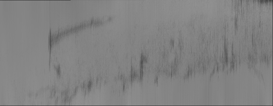
 Map data Map data | Reconstructed tomogram DS3-TS1 | ||||||||||||||||||
 Sample Sample |
| ||||||||||||||||||
 Keywords Keywords |  Amyloid / amyloid beta / alzheimers / neurodegeneration / plaque / tomography / in situ / brain / cryoCLEM / pathology / PROTEIN FIBRIL Amyloid / amyloid beta / alzheimers / neurodegeneration / plaque / tomography / in situ / brain / cryoCLEM / pathology / PROTEIN FIBRIL | ||||||||||||||||||
| Biological species |  Mus musculus (house mouse) Mus musculus (house mouse) | ||||||||||||||||||
| Method | electron tomography / cryo EM | ||||||||||||||||||
 Authors Authors | Leistner C / Wilkinson M / Burgess A / Lovatt M / Goodbody S / Xu Y / Deuchars S / Radford SE / Ranson NA / Frank RAW | ||||||||||||||||||
| Funding support |  United Kingdom, 5 items United Kingdom, 5 items
| ||||||||||||||||||
 Citation Citation | Journal: Nat Commun / Year: 2023 Title: The in-tissue molecular architecture of β-amyloid pathology in the mammalian brain. Authors: Conny Leistner / Martin Wilkinson / Ailidh Burgess / Megan Lovatt / Stanley Goodbody / Yong Xu / Susan Deuchars / Sheena E Radford / Neil A Ranson / René A W Frank / Abstract: Amyloid plaques composed of Aβ fibrils are a hallmark of Alzheimer's disease (AD). However, the molecular architecture of amyloid plaques in the context of fresh mammalian brain tissue is unknown. ...Amyloid plaques composed of Aβ fibrils are a hallmark of Alzheimer's disease (AD). However, the molecular architecture of amyloid plaques in the context of fresh mammalian brain tissue is unknown. Here, using cryogenic correlated light and electron tomography we report the in situ molecular architecture of Aβ fibrils in the App familial AD mouse model containing the Arctic mutation and an atomic model of ex vivo purified Arctic Aβ fibrils. We show that in-tissue Aβ fibrils are arranged in a lattice or parallel bundles, and are interdigitated by subcellular compartments, extracellular vesicles, extracellular droplets and extracellular multilamellar bodies. The Arctic Aβ fibril differs significantly from an earlier App fibril structure, indicating a striking effect of the Arctic mutation. These structural data also revealed an ensemble of additional fibrillar species, including thin protofilament-like rods and branched fibrils. Together, these results provide a structural model for the dense network architecture that characterises β-amyloid plaque pathology. | ||||||||||||||||||
| History |
|
- Structure visualization
| Supplemental images |
|---|

- Downloads & links
-EMDB archive
| Map data | emd_18301.map.gz | 171.4 MB |  EMDB map data format EMDB map data format | |
|---|---|---|---|---|
| Header (meta data) | emd-18301-v30.xmlemd-18301.xml | 12.7 KB 12.7 KB | Display Display | EMDB header |
| Images | 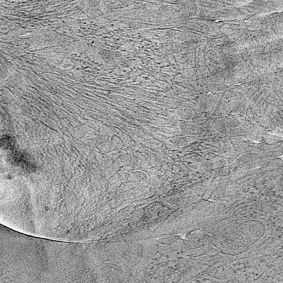 emd_18301.png emd_18301.png | 236.1 KB | ||
| Others | emd_18301_additional_1.map.gz | 200.5 MB | ||
| Archive directory |  http://ftp.pdbj.org/pub/emdb/structures/EMD-18301ftp://ftp.pdbj.org/pub/emdb/structures/EMD-18301 http://ftp.pdbj.org/pub/emdb/structures/EMD-18301ftp://ftp.pdbj.org/pub/emdb/structures/EMD-18301 | HTTPS FTP |
-Related structure data


-Links
| EMDB pages | EMDB (EBI/PDBe) / EMDataResource |
|---|
-Map
| File | Download / File: emd_18301.map.gz / Format: CCP4 / Size: 252.3 MB / Type: IMAGE STORED AS SIGNED BYTE | ||||||||||||||||||||
|---|---|---|---|---|---|---|---|---|---|---|---|---|---|---|---|---|---|---|---|---|---|
| Annotation | Reconstructed tomogram DS3-TS1 | ||||||||||||||||||||
| Voxel size | X=Y=Z: 13.7 Å | ||||||||||||||||||||
| Density |
| ||||||||||||||||||||
| Symmetry | Space group: 1 | ||||||||||||||||||||
| Details | EMDB XML:
|
-Supplemental data
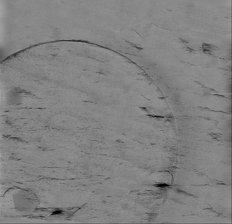
-Additional map: Reconstructed tomogram DS3-TS4
| File | emd_18301_additional_1.map | ||||||||||||
|---|---|---|---|---|---|---|---|---|---|---|---|---|---|
| Annotation | Reconstructed tomogram DS3-TS4 | ||||||||||||
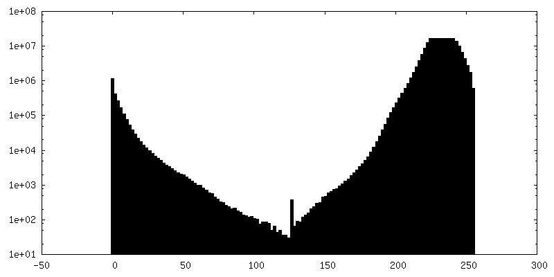
| Projections & Slices |
| ||||||||||||
| Density Histograms |
 Z
Z Y
Y X
X


- Sample components
-Entire : High-pressure frozen App^NL-G-F brain tissue containing Arctic am...
| Entire | Name: High-pressure frozen App^NL-G-F brain tissue containing Arctic amyloid-beta plaques |
|---|---|
| Components |
|
-Supramolecule #1: High-pressure frozen App^NL-G-F brain tissue containing Arctic am...
| Supramolecule | Name: High-pressure frozen App^NL-G-F brain tissue containing Arctic amyloid-beta plaques type: tissue / ID: 1 / Parent: 0 |
|---|---|
| Source (natural) | Organism: Mus musculus (house mouse) / Strain: App^NL-G-F / Organ: Brain |
-Experimental details
-Structure determination
| Method | cryo EM |
|---|---|
 Processing Processing | electron tomography |
| Aggregation state | tissue |
-Sample preparation
| Buffer | pH: 7.4 |
|---|---|
| Grid | Model: Quantifoil R1.2/1.3 / Material: COPPER / Mesh: 300 / Pretreatment - Type: GLOW DISCHARGE / Pretreatment - Time: 60 sec. |
| Vitrification | Cryogen name: ETHANE |
| High pressure freezing | Instrument: OTHER Details: 2000 bar. The value given for _em_high_pressure_freezing.instrument is Leica EM ICE. This is not in a list of allowed values {'OTHER', 'EMS-002 RAPID IMMERSION FREEZER', 'LEICA EM PACT2', ...Details: 2000 bar. The value given for _em_high_pressure_freezing.instrument is Leica EM ICE. This is not in a list of allowed values {'OTHER', 'EMS-002 RAPID IMMERSION FREEZER', 'LEICA EM PACT2', 'LEICA EM HPM100', 'LEICA EM PACT', 'BAL-TEC HPM 010'} so OTHER is written into the XML file. |
| Cryo protectant | 20% Dextran 40000 |
| Sectioning | Ultramicrotomy - Instrument: cryo-ultramicrotome (Leica EM FC7) Ultramicrotomy - Temperature: 123 K / Ultramicrotomy - Final thickness: 100 |
- Electron microscopy
| Microscope | FEI TITAN KRIOS |
|---|---|
| Electron beam | Acceleration voltage: 300 kV / Electron source: FIELD EMISSION GUN |
| Electron optics | Illumination mode: FLOOD BEAM / Imaging mode: BRIGHT FIELDBright-field microscopy / Nominal defocus max: 5.0 µm / Nominal defocus min: 4.0 µm |
| Image recording | Film or detector model: GATAN K2 SUMMIT (4k x 4k) / Detector mode: COUNTING / Average exposure time: 2.0 sec. / Average electron dose: 61.0 e/Å2 |
| Experimental equipment |  Model: Titan Krios / Image courtesy: FEI Company |
-Image processing
| Final reconstruction | Software - Name: IMOD / Number images used: 61 |
|---|
