 Movie
Movie Controller

[English] 日本語
 Yorodumi
- EMDB-18239: Structure of the recycling U5 snRNP bound to chaperone CD2BP2 and... -
+ Open data

- Basic information
| Entry |  | |||||||||
|---|---|---|---|---|---|---|---|---|---|---|
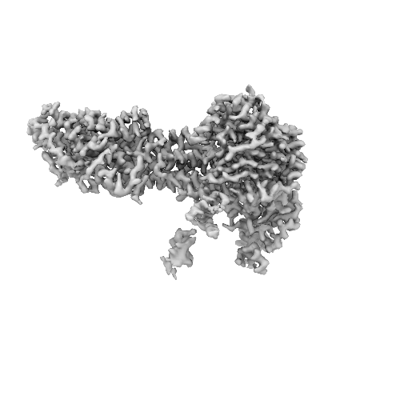
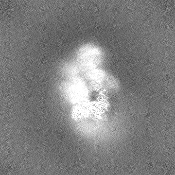
| Title | Structure of the recycling U5 snRNP bound to chaperone CD2BP2 and TSSC4 (Map 6) | |||||||||
 Map data Map data | sharpened map | |||||||||
 Sample Sample |
| |||||||||
 Keywords Keywords | U5 snRNP / CD2BP2 / TSSC4 /  spliceosome / splicing spliceosome / splicing | |||||||||
| Biological species |  Homo sapiens (human) Homo sapiens (human) | |||||||||
| Method | single particle reconstruction / cryo EM / Resolution: 2.7 Å | |||||||||
 Authors Authors | Riabov Bassat D / Plaschka C / Vorlaender MK | |||||||||
| Funding support | European Union, 2 items
| |||||||||
 Citation Citation | Journal: Nat Struct Mol Biol / Year: 2024 Title: Structural basis of human U5 snRNP late biogenesis and recycling. Authors: Daria Riabov Bassat / Supapat Visanpattanasin / Matthias K Vorländer / Laura Fin / Alexander W Phillips / Clemens Plaschka /  Abstract: Pre-mRNA splicing by the spliceosome requires the biogenesis and recycling of its small nuclear ribonucleoprotein (snRNP) complexes, which are consumed in each round of splicing. The human U5 snRNP ...Pre-mRNA splicing by the spliceosome requires the biogenesis and recycling of its small nuclear ribonucleoprotein (snRNP) complexes, which are consumed in each round of splicing. The human U5 snRNP is the ~1 MDa 'heart' of the spliceosome and is recycled through an unknown mechanism involving major architectural rearrangements and the dedicated chaperones CD2BP2 and TSSC4. Late steps in U5 snRNP biogenesis similarly involve these chaperones. Here we report cryo-electron microscopy structures of four human U5 snRNP-CD2BP2-TSSC4 complexes, revealing how a series of molecular events primes the U5 snRNP to generate the ~2 MDa U4/U6.U5 tri-snRNP, the largest building block of the spliceosome. | |||||||||
| History |
|
- Structure visualization
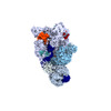
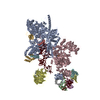
| Supplemental images |
|---|
- Downloads & links
-EMDB archive
| Map data | emd_18239.map.gz | 168.1 MB |  EMDB map data format EMDB map data format | |
|---|---|---|---|---|
| Header (meta data) | emd-18239-v30.xmlemd-18239.xml | 16.5 KB 16.5 KB | Display Display | EMDB header |
| FSC (resolution estimation) | emd_18239_fsc.xml | 11.8 KB | Display | FSC data file |
| Images |  emd_18239.png emd_18239.png | 49.3 KB | ||
| Filedesc metadata | emd-18239.cif.gz | 3.9 KB | ||
| Others | emd_18239_additional_1.map.gzemd_18239_half_map_1.map.gzemd_18239_half_map_2.map.gz | 90.2 MB 164.9 MB 164.9 MB | ||
| Archive directory |  http://ftp.pdbj.org/pub/emdb/structures/EMD-18239ftp://ftp.pdbj.org/pub/emdb/structures/EMD-18239 http://ftp.pdbj.org/pub/emdb/structures/EMD-18239ftp://ftp.pdbj.org/pub/emdb/structures/EMD-18239 | HTTPS FTP |
-Related structure data


-Links
| EMDB pages | EMDB (EBI/PDBe) / EMDataResource |
|---|
-Map
| File | Download / File: emd_18239.map.gz / Format: CCP4 / Size: 178 MB / Type: IMAGE STORED AS FLOATING POINT NUMBER (4 BYTES) | ||||||||||||||||||||
|---|---|---|---|---|---|---|---|---|---|---|---|---|---|---|---|---|---|---|---|---|---|
| Annotation | sharpened map | ||||||||||||||||||||
| Voxel size | X=Y=Z: 1.24 Å | ||||||||||||||||||||
| Density |
| ||||||||||||||||||||
| Symmetry | Space group: 1 | ||||||||||||||||||||
| Details | EMDB XML:
|
-Supplemental data
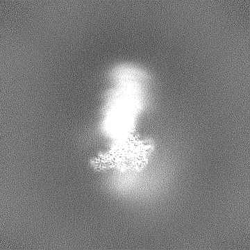
-Additional map: unsharpened map
| File | emd_18239_additional_1.map | ||||||||||||
|---|---|---|---|---|---|---|---|---|---|---|---|---|---|
| Annotation | unsharpened map | ||||||||||||
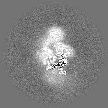
| Projections & Slices |
| ||||||||||||
| Density Histograms |
 Z
Z Y
Y X
X


-Half map: half map 2
| File | emd_18239_half_map_1.map | ||||||||||||
|---|---|---|---|---|---|---|---|---|---|---|---|---|---|
| Annotation | half map 2 | ||||||||||||
| Projections & Slices |
| ||||||||||||
| Density Histograms |


-Half map: half map 1
| File | emd_18239_half_map_2.map | ||||||||||||
|---|---|---|---|---|---|---|---|---|---|---|---|---|---|
| Annotation | half map 1 | ||||||||||||
| Projections & Slices |
| ||||||||||||
| Density Histograms |


- Sample components
-Entire : Structure of the recycling U5 snRNP bound to chaperone CD2BP2 and...
| Entire | Name: Structure of the recycling U5 snRNP bound to chaperone CD2BP2 and TSSC4 (Map 6) |
|---|---|
| Components |
|
-Supramolecule #1: Structure of the recycling U5 snRNP bound to chaperone CD2BP2 and...
| Supramolecule | Name: Structure of the recycling U5 snRNP bound to chaperone CD2BP2 and TSSC4 (Map 6) type: complex / ID: 1 / Parent: 0 / Macromolecule list: #1-#15 |
|---|---|
| Source (natural) | Organism: Homo sapiens (human) |
| Molecular weight | Theoretical: 1 MDa |
-Experimental details
-Structure determination
| Method | cryo EM |
|---|---|
 Processing Processing | single particle reconstruction |
| Aggregation state | particle |
-Sample preparation
| Buffer | pH: 7.9 |
|---|---|
| Vitrification | Cryogen name: ETHANE |
- Electron microscopy
| Microscope | FEI TITAN KRIOS |
|---|---|
| Electron beam | Acceleration voltage: 300 kV / Electron source: FIELD EMISSION GUN |
| Electron optics | Illumination mode: FLOOD BEAM / Imaging mode: BRIGHT FIELDBright-field microscopy / Nominal defocus max: 2.0 µm / Nominal defocus min: 0.5 µm |
| Image recording | Film or detector model: FEI FALCON IV (4k x 4k) / Average electron dose: 40.0 e/Å2 |
| Experimental equipment |  Model: Titan Krios / Image courtesy: FEI Company |
-Image processing
| Startup model | Type of model: PDB ENTRY PDB model - PDB ID: |
|---|---|
| Initial angle assignment | Type: MAXIMUM LIKELIHOOD |
| Final angle assignment | Type: MAXIMUM LIKELIHOOD |
| Final reconstruction | Resolution.type: BY AUTHOR / Resolution: 2.7 Å / Resolution method: FSC 0.143 CUT-OFF / Number images used: 256427 |
| FSC plot (resolution estimation) |  |

