 Movie
Movie Controller

[English] 日本語
 Yorodumi
- EMDB-18201: E. coli plasmid-borne JetABCD(E248A) core in a cleavage-competent... -
+ Open data

- Basic information
| Entry | 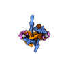 | |||||||||
|---|---|---|---|---|---|---|---|---|---|---|
| Title | E. coli plasmid-borne JetABCD(E248A) core in a cleavage-competent state | |||||||||
 Map data Map data | ||||||||||
 Sample Sample |
| |||||||||
 Keywords Keywords | SMC complexes / DNA loop extrusion /  nuclease / bacterial immunity / defense system / topoisomerase / plamid restriction / MksBEFG / Wadjet / EptABCD / horizontal gene transfer / DNA binding protein / DNA kinking nuclease / bacterial immunity / defense system / topoisomerase / plamid restriction / MksBEFG / Wadjet / EptABCD / horizontal gene transfer / DNA binding protein / DNA kinking | |||||||||
| Function / homology |  Function and homology information Function and homology informationP-loop containing region of AAA domain / Uncharacterised conserved protein UCP028408 / Wadjet protein JetD, C-terminal / Domain of unknown function DUF3322 / Wadjet protein JetD, C-terminal / Uncharacterized protein conserved in bacteria N-term (DUF3322) / Protein of unknown function DUF3375 / Protein of unknown function (DUF3375) / SbcC/RAD50-like, Walker B motif / P-loop containing nucleoside triphosphate hydrolaseSimilarity search - Domain/homology | |||||||||
| Biological species |  Escherichia coli (E. coli) Escherichia coli (E. coli) | |||||||||
| Method | single particle reconstruction / cryo EM / Resolution: 4.17 Å | |||||||||
 Authors Authors | Roisne-Hamelin F / Li Y / Gruber S | |||||||||
| Funding support | European Union, 1 items
| |||||||||
 Citation Citation | Journal: Mol Cell / Year: 2024 Title: Structural basis for plasmid restriction by SMC JET nuclease. Authors: Florian Roisné-Hamelin / Hon Wing Liu / Michael Taschner / Yan Li / Stephan Gruber /  Abstract: DNA loop-extruding SMC complexes play crucial roles in chromosome folding and DNA immunity. Prokaryotic SMC Wadjet (JET) complexes limit the spread of plasmids through DNA cleavage, yet the ...DNA loop-extruding SMC complexes play crucial roles in chromosome folding and DNA immunity. Prokaryotic SMC Wadjet (JET) complexes limit the spread of plasmids through DNA cleavage, yet the mechanisms for plasmid recognition are unresolved. We show that artificial DNA circularization renders linear DNA susceptible to JET nuclease cleavage. Unlike free DNA, JET cleaves immobilized plasmid DNA at a specific site, the plasmid-anchoring point, showing that the anchor hinders DNA extrusion but not DNA cleavage. Structures of plasmid-bound JetABC reveal two presumably stalled SMC motor units that are drastically rearranged from the resting state, together entrapping a U-shaped DNA segment, which is further converted to kinked V-shaped cleavage substrate by JetD nuclease binding. Our findings uncover mechanical bending of residual unextruded DNA as molecular signature for plasmid recognition and non-self DNA elimination. We moreover elucidate key elements of SMC loop extrusion, including the motor direction and the structure of a DNA-holding state. | |||||||||
| History |
|
- Structure visualization
| Supplemental images |
|---|

- Downloads & links
-EMDB archive
| Map data | emd_18201.map.gz | 118.2 MB | EMDB map data format | |
|---|---|---|---|---|
| Header (meta data) | emd-18201-v30.xmlemd-18201.xml | 25.7 KB 25.7 KB | Display Display | EMDB header |
| Images |  emd_18201.png emd_18201.png | 47 KB | ||
| Masks | emd_18201_msk_1.map | 125 MB | Mask map | |
| Filedesc metadata | emd-18201.cif.gz | 8.1 KB | ||
| Others | emd_18201_additional_1.map.gzemd_18201_half_map_1.map.gzemd_18201_half_map_2.map.gz | 63 MB 115.9 MB 115.9 MB | ||
| Archive directory |  http://ftp.pdbj.org/pub/emdb/structures/EMD-18201ftp://ftp.pdbj.org/pub/emdb/structures/EMD-18201 http://ftp.pdbj.org/pub/emdb/structures/EMD-18201ftp://ftp.pdbj.org/pub/emdb/structures/EMD-18201 | HTTPS FTP |
-Related structure data
| Related structure data | 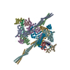 8q72MC C: citing same article ( M: atomic model generated by this map |
|---|---|
| Similar structure data |


-Links
| EMDB pages | EMDB (EBI/PDBe) / EMDataResource |
|---|
-Map
| File | Download / File: emd_18201.map.gz / Format: CCP4 / Size: 125 MB / Type: IMAGE STORED AS FLOATING POINT NUMBER (4 BYTES) | ||||||||||||||||||||
|---|---|---|---|---|---|---|---|---|---|---|---|---|---|---|---|---|---|---|---|---|---|
| Voxel size | X=Y=Z: 1.66 Å | ||||||||||||||||||||
| Density |
| ||||||||||||||||||||
| Symmetry | Space group: 1 | ||||||||||||||||||||
| Details | EMDB XML:
|
-Supplemental data
-Mask #1
| File | emd_18201_msk_1.map | ||||||||||||
|---|---|---|---|---|---|---|---|---|---|---|---|---|---|
| Projections & Slices |
| ||||||||||||
| Density Histograms |
 Z
Z Y
Y X
X


-Additional map: This is the unsharpened map.
| File | emd_18201_additional_1.map | ||||||||||||
|---|---|---|---|---|---|---|---|---|---|---|---|---|---|
| Annotation | This is the unsharpened map. | ||||||||||||
| Projections & Slices |
| ||||||||||||
| Density Histograms |


-Half map: #2
| File | emd_18201_half_map_1.map | ||||||||||||
|---|---|---|---|---|---|---|---|---|---|---|---|---|---|
| Projections & Slices |
| ||||||||||||
| Density Histograms |


-Half map: #1
| File | emd_18201_half_map_2.map | ||||||||||||
|---|---|---|---|---|---|---|---|---|---|---|---|---|---|
| Projections & Slices |
| ||||||||||||
| Density Histograms |


- Sample components
-Entire : JetABCD(E248A) loaded onto plasmid DNA
| Entire | Name: JetABCD(E248A) loaded onto plasmid DNA |
|---|---|
| Components |
|
-Supramolecule #1: JetABCD(E248A) loaded onto plasmid DNA
| Supramolecule | Name: JetABCD(E248A) loaded onto plasmid DNA / type: complex / ID: 1 / Parent: 0 / Macromolecule list: #1-#5 Details: E. coli JetABC, JetD(E248A) and plasmid DNA were mixed and incubated with ATP prior freezing. |
|---|---|
| Source (natural) | Organism: Escherichia coli (E. coli) / Strain: GF4-3 |
-Macromolecule #1: JetC
| Macromolecule | Name: JetC / type: protein_or_peptide / ID: 1 Details: The last "G" in the theorical sequence is the result of a DNA cloning scar. Number of copies: 4 / Enantiomer: LEVO |
|---|---|
| Source (natural) | Organism: Escherichia coli (E. coli) / Strain: GF4-3 |
| Molecular weight | Theoretical: 124.562938 KDa |
| Recombinant expression | Organism: Escherichia coli (E. coli) |
| Sequence | String: MNQVSGLAGK ESFILTRIEL FNWGGFHGLH QAAIHQDGTA VIGPTGSGKT TLVDALMTLL CANPRYNLAS TGGHESDRDL ISYVRGVSG PGDGGEGQSH IARPGKTVTG IAATLEREGK QVRLGALLWF DSTSSSVTDM KRLWLFSDNP GQTLEHWLNV Y HEGGTRLL ...String: MNQVSGLAGK ESFILTRIEL FNWGGFHGLH QAAIHQDGTA VIGPTGSGKT TLVDALMTLL CANPRYNLAS TGGHESDRDL ISYVRGVSG PGDGGEGQSH IARPGKTVTG IAATLEREGK QVRLGALLWF DSTSSSVTDM KRLWLFSDNP GQTLEHWLNV Y HEGGTRLL RQMEKEAIGL WTYPNKKQYL ARLRDFFEVG ENAFTLLNRA AGLKQLNSID EIFRELVLDD HSAFDRAAEV AN SFDGLTE IHQELETARK QQQSLQPVAL SWEKYQKQER QLADWLTLES LLPLWFAQQA SHLWREKINL LNARLAEAQT SEE QLQSQL DLQKKVVSDC MQRYLQVGGA NIDELNERIK DWQKTLGSRE ALARQYQQLT RNLGLPSDLS QPQLEANQHE AEAR CEQIA VDIKLKQEEA YQKGALSHHI TEELRERENE RAEIARRPDS NLPAHYQAFR SELAKALNVD ESELPFVAEL IQVKP EEAQ WRGAIERAVG SNRLRILVAP ESAQEALRWV NQRNNRLHVR LLEVKLPHSP ARFFDDGFTR KLLWKDHPWR EAVKAL LAE SDRHCVDSPE QLHDTPHAMT VQGLMSGKQR FYDKHDQKRL DEDWLTGFDN RDRLNFLAKE IATLQEQVKT ANAAFEF AK GEVGLLQNQA ASFQKIEQID FDSIDVPGAK SQLDALRERL ENLTRPDSDA SVAKAKLDEA QTIESELDKQ LRAANKVT N VLDTELTLAR AAERKAQQTA QQGMKEEERE LCASHFPVVT LEQLPDIRDL ERQHERGIQH EIERVKAELH RLNIELTKR MSEAKRVDTG ALVEAGADLD DIPVYLQRLQ ELTEEALPEK LNRFLDYLNR SSDDGVTQLL SHIEHEVLVI EERLNELNET MFRVDFQPD RYLRLDTKKV VHESLRTLEK AQRQLNAARF VDDNGESHYK ALQVLVAQLR DACERNRTLG AKALLDPRFR L EFAVSVMD RQSGNVIESR TGSQGGSGGE KEIIASYVLT ASLSYALCPA GSRYPLFGTI ILDEAFSRSS HAVAGRIIAA LR EFGLHAV FITPNKEMRL LRDHTRSAIV VHRRGQNSNM ASLSWEELER HYQRRGNAG UniProtKB: Chromosome partition protein Smc |
-Macromolecule #2: JetB
| Macromolecule | Name: JetB / type: protein_or_peptide / ID: 2 Details: The last "G" in the theorical sequence is the result of a DNA cloning scar. Number of copies: 4 / Enantiomer: LEVO |
|---|---|
| Source (natural) | Organism: Escherichia coli (E. coli) / Strain: GF4-3 |
| Molecular weight | Theoretical: 28.020416 KDa |
| Recombinant expression | Organism: Escherichia coli (E. coli) |
| Sequence | String: MAGFFDKLIN RSVTANAGCE PEPSDEEVTD ESVEDSLASS ETRTLQKIRE ATQELLKYGL LEEASKPNLY RIVLSHPEEV TRILEPLDL DIGIDEIRGL LYVKVRLDET PAQDEWAHPL VRRQRLNLEQ SLLVAILRQH FVAWEQESGT GASQAQIAID D LLPQLQIY ...String: MAGFFDKLIN RSVTANAGCE PEPSDEEVTD ESVEDSLASS ETRTLQKIRE ATQELLKYGL LEEASKPNLY RIVLSHPEEV TRILEPLDL DIGIDEIRGL LYVKVRLDET PAQDEWAHPL VRRQRLNLEQ SLLVAILRQH FVAWEQESGT GASQAQIAID D LLPQLQIY LGDPGSESKE RTRLLTLLDQ LKGHGLVTSP DAHERIVIRP IIAHLADPIN LQALLAWLRE QIAQQTSPND AP EKDSSEE DVG |
-Macromolecule #3: JetA
| Macromolecule | Name: JetA / type: protein_or_peptide / ID: 3 Details: "GPAA" at the begining of the theorical sequence is the remaining of the purification tag after tag cleavage. The last "G" in the theorical sequence is the result of a DNA cloning scar. Number of copies: 2 / Enantiomer: LEVO |
|---|---|
| Source (natural) | Organism: Escherichia coli (E. coli) / Strain: GF4-3 |
| Molecular weight | Theoretical: 57.81891 KDa |
| Recombinant expression | Organism: Escherichia coli (E. coli) |
| Sequence | String: GPAAMEENTR QRTENYISAK NQHPAWILLA TRRAPLVLSC LKTLFEKSHD GIPLEEAIQS LSSILIEHVS QEQYDINQDN PFLQASREL REWIKRRLIV ERDGRIFATD ALEVAITFVE SLDNRFMTST ASRLSTVQRE IENLETRLNP NPANRVATLR R RISELERE ...String: GPAAMEENTR QRTENYISAK NQHPAWILLA TRRAPLVLSC LKTLFEKSHD GIPLEEAIQS LSSILIEHVS QEQYDINQDN PFLQASREL REWIKRRLIV ERDGRIFATD ALEVAITFVE SLDNRFMTST ASRLSTVQRE IENLETRLNP NPANRVATLR R RISELERE LQEAEAGHIE VLETHQAVEH IRDVYNLASS LRADFRRVED SWREADRALR QSIIGEQYHR GDIVERLLND QD ALLNTPE GRVFDSFQQQ LRQSSELKAM SERLRVILSH PSASDALNRL QRHDLRWLVK RLVDESQTVL QARARSERDV RGF MKTGLA AEHHRVGHLL NEFLNLALKL DWQRQMIRKQ EVPLPAVGVA VTGIPAIERL RFKEVDDEAE QTLDLSNHAA DLTQ IGDDF WDAFNGLDRE VLIQQTLQLL AKENRPVGLA ELAELLPPAH DLETFAVWIG MAREAGIEVI DSQREFAELS DGEGR RWRF NLPTTGLESQ ALMDIDWEG UniProtKB: DUF3375 family protein |
-Macromolecule #4: JetD(E248A)
| Macromolecule | Name: JetD(E248A) / type: protein_or_peptide / ID: 4 Details: "GSLEVLFQ" at the C-terminus in the theorical sequence is the remaining of the tag after tag cleavage. The sequence carries the "E248A" mutation (nuclease dead). Number of copies: 2 / Enantiomer: LEVO |
|---|---|
| Source (natural) | Organism: Escherichia coli (E. coli) |
| Molecular weight | Theoretical: 43.938492 KDa |
| Recombinant expression | Organism: Escherichia coli (E. coli) |
| Sequence | String: MYSPDELREK LARQWDSAKL RAERLLSPGN WPLCLPIGKP STKIFAEQTQ RVLQHVQRWR QVAVGHVEWE AVSFRASDTP VLMPLRWIL NSPSEWINAA ADPVVSREFR LLEGIIEQVN PIFHPLLVKH RSLWRHKDPQ GVISAATLAC RLEPGCAKGL P LRLLSGQG ...String: MYSPDELREK LARQWDSAKL RAERLLSPGN WPLCLPIGKP STKIFAEQTQ RVLQHVQRWR QVAVGHVEWE AVSFRASDTP VLMPLRWIL NSPSEWINAA ADPVVSREFR LLEGIIEQVN PIFHPLLVKH RSLWRHKDPQ GVISAATLAC RLEPGCAKGL P LRLLSGQG VDTKFIENNI SLLTRLLDVR FSGEASEQGL TTFLDAFDES SHWVLVVPLS PGLLPFKKCR VTTAELAETT LP ASQVLVV ANEQCLHHLP ALSDTIAILG CGLDVQWLSS SVLDEKRVAY WGDMDSWGLL MLARARRCRP TLDALLMNRE LFE QYASHS AVPEPVIAKE AVPDGLLNEE ADFYRYLTRL PRGRLEQEFL PVGIAEEALL RWGKESGSLE VLFQ UniProtKB: DUF3322 and DUF2220 domain-containing protein |
-Macromolecule #5: Circular plasmid DNA (1840-MER)
| Macromolecule | Name: Circular plasmid DNA (1840-MER) / type: dna / ID: 5 Details: Only a portion of the plasmid (pSG6085, 1840 bp) has been modelled as polyAT track, because the complex is expected to bind in random position to the DNA and the resolution of DNA do not ...Details: Only a portion of the plasmid (pSG6085, 1840 bp) has been modelled as polyAT track, because the complex is expected to bind in random position to the DNA and the resolution of DNA do not allow any sequence assignement. Number of copies: 4 / Classification: DNA |
|---|---|
| Source (natural) | Organism: Escherichia coli (E. coli) |
| Molecular weight | Theoretical: 12.303033 KDa |
| Sequence | String: (DA)(DT)(DA)(DT)(DA)(DT)(DA)(DT)(DA)(DT) (DA)(DT)(DA)(DT)(DA)(DT)(DA)(DT)(DA)(DT) (DA)(DT)(DA)(DT)(DA)(DT)(DA)(DT)(DA) (DT)(DA)(DT)(DA)(DT)(DA)(DT)(DA)(DT)(DA) (DT) |
-Macromolecule #6: ADENOSINE-5'-DIPHOSPHATE
| Macromolecule | Name: ADENOSINE-5'-DIPHOSPHATE / type: ligand / ID: 6 / Number of copies: 4 / Formula: ADP |
|---|---|
| Molecular weight | Theoretical: 427.201 Da |
| Chemical component information |  ChemComp-ADP: |
-Experimental details
-Structure determination
| Method | cryo EM |
|---|---|
 Processing Processing | single particle reconstruction |
| Aggregation state | particle |
-Sample preparation
| Buffer | pH: 7.5 |
|---|---|
| Grid | Model: Au-flat 1.2/1.3 / Material: GOLD / Pretreatment - Type: GLOW DISCHARGE |
| Vitrification | Cryogen name: ETHANE / Chamber humidity: 100 % / Chamber temperature: 283 K / Instrument: FEI VITROBOT MARK IV |
- Electron microscopy
| Microscope | TFS KRIOS |
|---|---|
| Electron beam | Acceleration voltage: 300 kV / Electron source: FIELD EMISSION GUN |
| Electron optics | Illumination mode: FLOOD BEAM / Imaging mode: BRIGHT FIELDBright-field microscopy / Nominal defocus max: 2.6 µm / Nominal defocus min: 0.8 µm |
| Image recording | Film or detector model: FEI FALCON IV (4k x 4k) / Number grids imaged: 1 / Number real images: 37902 / Average electron dose: 40.0 e/Å2 |
| Experimental equipment |  Model: Titan Krios / Image courtesy: FEI Company |
-Image processing
| Startup model | Type of model: INSILICO MODEL |
|---|---|
| Initial angle assignment | Type: MAXIMUM LIKELIHOOD |
| Final angle assignment | Type: MAXIMUM LIKELIHOOD |
| Final reconstruction | Applied symmetry - Point group: C2 (2 fold cyclic) / Resolution.type: BY AUTHOR / Resolution: 4.17 Å / Resolution method: FSC 0.143 CUT-OFF / Software - Name: cryoSPARC (ver. 3.3) / Number images used: 235956 |
-Atomic model buiding 1
| Initial model |
| ||||||||||||
|---|---|---|---|---|---|---|---|---|---|---|---|---|---|
| Refinement | Protocol: FLEXIBLE FIT | ||||||||||||
| Output model | PDB-8q72: |

