 Movie
Movie Controller

[English] 日本語
 Yorodumi
- EMDB-17823: Tomogram of the Emiliania huxleyi virus 201 (EhV-201) purified pa... -
+ Open data

- Basic information
| Entry |  | |||||||||
|---|---|---|---|---|---|---|---|---|---|---|
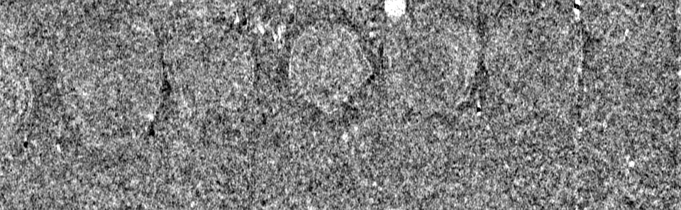
| Title | Tomogram of the Emiliania huxleyi virus 201 (EhV-201) purified particles used for subtomogram averaging. | |||||||||
 Map data Map data | Original tomogram | |||||||||
 Sample Sample |
| |||||||||
 Keywords Keywords |  cryo-EM / subtomogram averaging / EhV-201 / enveloped virus / capsid / major capsid protein / VIRUS cryo-EM / subtomogram averaging / EhV-201 / enveloped virus / capsid / major capsid protein / VIRUS | |||||||||
| Biological species |  Emiliania huxleyi virus 201 Emiliania huxleyi virus 201 | |||||||||
| Method | electron tomography / cryo EM / Resolution: 25.0 Å | |||||||||
 Authors Authors | Homola M / Buttner CR / Fuzik T / Novacek J / Chaillet M / Forster F / Plevka P | |||||||||
| Funding support |  Czech Republic, European Union, 2 items Czech Republic, European Union, 2 items
| |||||||||
 Citation Citation | Journal: To Be Published Title: Structure and replication cycle of a virus infecting climate-modulating alga Emiliania huxleyi Authors: Homola M / Buttner CR / Fuzik T / Novacek J / Chaillet M / Forster F / Plevka P | |||||||||
| History |
|
- Structure visualization
| Supplemental images |
|---|

- Downloads & links
-EMDB archive
| Map data | emd_17823.map.gz | 683.5 MB |  EMDB map data format EMDB map data format | |
|---|---|---|---|---|
| Header (meta data) | emd-17823-v30.xmlemd-17823.xml | 14.4 KB 14.4 KB | Display Display | EMDB header |
| Images | 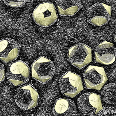 emd_17823.png emd_17823.png | 319.2 KB | ||
| Others | emd_17823_additional_1.map.gz | 149.1 MB | ||
| Archive directory |  http://ftp.pdbj.org/pub/emdb/structures/EMD-17823ftp://ftp.pdbj.org/pub/emdb/structures/EMD-17823 http://ftp.pdbj.org/pub/emdb/structures/EMD-17823ftp://ftp.pdbj.org/pub/emdb/structures/EMD-17823 | HTTPS FTP |
-Related structure data
| Related structure data |
|---|
-Links
| EMDB pages | EMDB (EBI/PDBe) / EMDataResource |
|---|
-Map
| File | Download / File: emd_17823.map.gz / Format: CCP4 / Size: 739.3 MB / Type: IMAGE STORED AS FLOATING POINT NUMBER (4 BYTES) | ||||||||||||||||||||||||||||||||
|---|---|---|---|---|---|---|---|---|---|---|---|---|---|---|---|---|---|---|---|---|---|---|---|---|---|---|---|---|---|---|---|---|---|
| Annotation | Original tomogram | ||||||||||||||||||||||||||||||||
| Projections & slices | Image control
Images are generated by Spider. generated in cubic-lattice coordinate | ||||||||||||||||||||||||||||||||
| Voxel size | X=Y=Z: 12.48 Å | ||||||||||||||||||||||||||||||||
| Density |
| ||||||||||||||||||||||||||||||||
| Symmetry | Space group: 1 | ||||||||||||||||||||||||||||||||
| Details | EMDB XML:
|
 Z (Sec.)
 Y (Row.)
 X (Col.)


-Supplemental data
-Additional map: Place back of virion vertices using coordinates from 3D refinement.
| File | emd_17823_additional_1.map | ||||||||||||
|---|---|---|---|---|---|---|---|---|---|---|---|---|---|
| Annotation | Place back of virion vertices using coordinates from 3D refinement. | ||||||||||||
| Projections & Slices |
| ||||||||||||
| Density Histograms |


- Sample components
-Entire : Emiliania huxleyi virus 201
| Entire | Name: Emiliania huxleyi virus 201 |
|---|---|
| Components |
|
-Supramolecule #1: Emiliania huxleyi virus 201
| Supramolecule | Name: Emiliania huxleyi virus 201 / type: virus / ID: 1 / Parent: 0 / Macromolecule list: #1-#2 Details: EhV-201 was propagated on a non-calcifying Emiliania huxleyi strain (CCPM 2090). NCBI-ID: 181210 / Sci species name: Emiliania huxleyi virus 201 / Virus type: VIRION / Virus isolate: SPECIES / Virus enveloped: Yes / Virus empty: No |
|---|---|
| Host (natural) | Organism:  Emiliania huxleyi CCMP1516 (eukaryote) / Strain: CCMP 2090 Emiliania huxleyi CCMP1516 (eukaryote) / Strain: CCMP 2090 |
| Virus shell | Shell ID: 1 / Name: inner membrane |
| Virus shell | Shell ID: 2 / Name: capsid / Diameter: 1990.0 Å / T number (triangulation number): 169 |
| Virus shell | Shell ID: 3 / Name: outer membrane / Diameter: 2110.0 Å |
-Experimental details
-Structure determination
| Method | cryo EM |
|---|---|
 Processing Processing | electron tomography |
| Aggregation state | particle |
-Sample preparation
| Buffer | pH: 8 / Component - Name: sea salt |
|---|---|
| Grid | Model: Quantifoil R2/1 / Material: COPPER / Mesh: 200 / Support film - Material: CARBON / Support film - topology: HOLEY / Support film - Film thickness: 12 / Pretreatment - Type: GLOW DISCHARGE / Pretreatment - Time: 15 sec. / Pretreatment - Atmosphere: OTHER / Pretreatment - Pressure: 5e-05 kPa / Details: top side only |
| Vitrification | Cryogen name: ETHANE / Chamber humidity: 100 % / Chamber temperature: 283.15 K / Instrument: FEI VITROBOT MARK IV Details: Sample: 3.5 ul; Wait time: 10 s; Blot time: 3 s; Blot force: -2; Drain time: 0 s. |
| Details | The viral sample was concentrated down to 1x10^10 plaque-forming units per ml (PFU ml^-1) |
| Sectioning | Other: NO SECTIONING |
| Fiducial marker | Manufacturer: Aurion / Diameter: 6 nm |
- Electron microscopy
| Microscope | FEI TITAN KRIOS |
|---|---|
| Electron beam | Acceleration voltage: 300 kV / Electron source: FIELD EMISSION GUN |
| Electron optics | C2 aperture diameter: 30.0 µm / Calibrated defocus max: 4.0 µm / Calibrated defocus min: 2.0 µm / Calibrated magnification: 42000 / Illumination mode: FLOOD BEAM / Imaging mode: BRIGHT FIELDBright-field microscopy / Cs: 2.7 mm / Nominal defocus max: 4.0 µm / Nominal defocus min: 2.0 µm / Nominal magnification: 42000 |
| Specialist optics | Energy filter - Name: GIF Bioquantum / Energy filter - Slit width: 10 eV |
| Sample stage | Specimen holder model: FEI TITAN KRIOS AUTOGRID HOLDER / Cooling holder cryogen: NITROGEN |
| Temperature | Min: 77.0 K / Max: 77.0 K |
| Image recording | Film or detector model: GATAN K3 BIOQUANTUM (6k x 4k) / Digitization - Dimensions - Width: 5760 pixel / Digitization - Dimensions - Height: 4092 pixel / Number real images: 4323 / Average exposure time: 1.5 sec. / Average electron dose: 2.42 e/Å2 |
| Experimental equipment |  Model: Titan Krios / Image courtesy: FEI Company |
-Image processing
| Final reconstruction | Algorithm: BACK PROJECTION / Resolution.type: BY AUTHOR / Resolution: 25.0 Å / Resolution method: OTHER / Number images used: 1 |
|---|
