 Movie
Movie Controller

[English] 日本語
 Yorodumi
- EMDB-16810: RcpA-TadD with C13 symmetry from the Pseudomonas aeruginosa Tight... -
+ Open data

- Basic information
| Entry |  | |||||||||
|---|---|---|---|---|---|---|---|---|---|---|
| Title | RcpA-TadD with C13 symmetry from the Pseudomonas aeruginosa Tight Adherence Secretion System | |||||||||
 Map data Map data | RcpA-TadD with C13 symmetry from the Pseudomonas aeruginosa Tigh Adherence Secretion System | |||||||||
 Sample Sample |
| |||||||||
 Keywords Keywords |  Secretin / Pilotin / RcpA / TadD / TAD / MEMBRANE PROTEIN Secretin / Pilotin / RcpA / TadD / TAD / MEMBRANE PROTEIN | |||||||||
| Function / homology |  Function and homology informationtype II protein secretion system complex / protein secretion / plasma membrane Function and homology informationtype II protein secretion system complex / protein secretion / plasma membraneSimilarity search - Function | |||||||||
| Biological species |  Pseudomonas aeruginosa PAO1 (bacteria) Pseudomonas aeruginosa PAO1 (bacteria) | |||||||||
| Method | single particle reconstruction / cryo EM / Resolution: 2.7 Å | |||||||||
 Authors Authors | Tassinari M / Low HH | |||||||||
| Funding support |  United Kingdom, 1 items United Kingdom, 1 items
| |||||||||
 Citation Citation | Journal: Nat Commun / Year: 2023 Title: Assembly mechanism of a Tad secretion system secretin-pilotin complex. Authors: Matteo Tassinari / Marta Rudzite / Alain Filloux / Harry H Low /   Abstract: The bacterial Tight adherence Secretion System (TadSS) assembles surface pili that drive cell adherence, biofilm formation and bacterial predation. The structure and mechanism of the TadSS is mostly ...The bacterial Tight adherence Secretion System (TadSS) assembles surface pili that drive cell adherence, biofilm formation and bacterial predation. The structure and mechanism of the TadSS is mostly unknown. This includes characterisation of the outer membrane secretin through which the pilus is channelled and recruitment of its pilotin. Here we investigate RcpA and TadD lipoprotein from Pseudomonas aeruginosa. Light microscopy reveals RcpA colocalising with TadD in P. aeruginosa and when heterologously expressed in Escherichia coli. We use cryogenic electron microscopy to determine how RcpA and TadD assemble a secretin channel with C13 and C14 symmetries. Despite low sequence homology, we show that TadD shares a similar fold to the type 4 pilus system pilotin PilF. We establish that the C-terminal four residues of RcpA bind TadD - an interaction essential for secretin formation. The binding mechanism between RcpA and TadD appears distinct from known secretin-pilotin pairings in other secretion systems. | |||||||||
| History |
|
- Structure visualization
| Supplemental images |
|---|

- Downloads & links
-EMDB archive
| Map data | emd_16810.map.gz | 6.9 MB | EMDB map data format | |
|---|---|---|---|---|
| Header (meta data) | emd-16810-v30.xmlemd-16810.xml | 17.2 KB 17.2 KB | Display Display | EMDB header |
| FSC (resolution estimation) | emd_16810_fsc.xml | 12.7 KB | Display | FSC data file |
| Images | 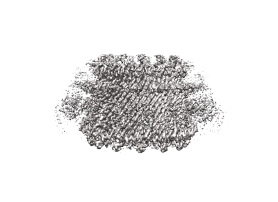 emd_16810.png emd_16810.png | 71.3 KB | ||
| Others | emd_16810_half_map_1.map.gzemd_16810_half_map_2.map.gz | 156.7 MB 135.3 MB | ||
| Archive directory |  http://ftp.pdbj.org/pub/emdb/structures/EMD-16810ftp://ftp.pdbj.org/pub/emdb/structures/EMD-16810 http://ftp.pdbj.org/pub/emdb/structures/EMD-16810ftp://ftp.pdbj.org/pub/emdb/structures/EMD-16810 | HTTPS FTP |
-Related structure data
| Related structure data |  8odnMC M: atomic model generated by this map C: citing same article ( |
|---|---|
| Similar structure data |

-Links
| EMDB pages | EMDB (EBI/PDBe) / EMDataResource |
|---|---|
| Related items in Molecule of the Month |


-Map
| File | Download / File: emd_16810.map.gz / Format: CCP4 / Size: 178 MB / Type: IMAGE STORED AS FLOATING POINT NUMBER (4 BYTES) | ||||||||||||||||||||
|---|---|---|---|---|---|---|---|---|---|---|---|---|---|---|---|---|---|---|---|---|---|
| Annotation | RcpA-TadD with C13 symmetry from the Pseudomonas aeruginosa Tigh Adherence Secretion System | ||||||||||||||||||||
| Voxel size | X=Y=Z: 1.1 Å | ||||||||||||||||||||
| Density |
| ||||||||||||||||||||
| Symmetry | Space group: 1 | ||||||||||||||||||||
| Details | EMDB XML:
|
-Supplemental data
-Half map: RcpA-TadD with C13 symmetry from the Pseudomonas aeruginosa...
| File | emd_16810_half_map_1.map | ||||||||||||
|---|---|---|---|---|---|---|---|---|---|---|---|---|---|
| Annotation | RcpA-TadD with C13 symmetry from the Pseudomonas aeruginosa Tigh Adherence Secretion System | ||||||||||||
| Projections & Slices |
| ||||||||||||
| Density Histograms |
 Z
Z Y
Y X
X


-Half map: RcpA-TadD with C13 symmetry from the Pseudomonas aeruginosa...
| File | emd_16810_half_map_2.map | ||||||||||||
|---|---|---|---|---|---|---|---|---|---|---|---|---|---|
| Annotation | RcpA-TadD with C13 symmetry from the Pseudomonas aeruginosa Tigh Adherence Secretion System | ||||||||||||
| Projections & Slices |
| ||||||||||||
| Density Histograms |


- Sample components
-Entire : RcpA-TadD with C13 symmetry
| Entire | Name: RcpA-TadD with C13 symmetry |
|---|---|
| Components |
|
-Supramolecule #1: RcpA-TadD with C13 symmetry
| Supramolecule | Name: RcpA-TadD with C13 symmetry / type: complex / ID: 1 / Parent: 0 / Macromolecule list: all |
|---|---|
| Source (natural) | Organism: Pseudomonas aeruginosa PAO1 (bacteria) |
| Molecular weight | Theoretical: 900 KDa |
-Macromolecule #1: RcpA
| Macromolecule | Name: RcpA / type: protein_or_peptide / ID: 1 / Number of copies: 13 / Enantiomer: LEVO |
|---|---|
| Source (natural) | Organism: Pseudomonas aeruginosa PAO1 (bacteria) |
| Molecular weight | Theoretical: 45.507402 KDa |
| Recombinant expression | Organism: Escherichia coli (E. coli) |
| Sequence | String: MHRSTGIGVS RWLGGLLGVA LALPALALPQ GCIELLAQAP RVDVVQGQQR DLRLAVPIER LAIGDPKIAD VQLLDRRGFL VTGKEQGST SLLIWTGCSP EPLRSLVEVE GRGSVDTRGA PAFTVGAAEE LPNQVQTDIR FVEVSRSKLK QASTSFVRRG G NLWVLGAP ...String: MHRSTGIGVS RWLGGLLGVA LALPALALPQ GCIELLAQAP RVDVVQGQQR DLRLAVPIER LAIGDPKIAD VQLLDRRGFL VTGKEQGST SLLIWTGCSP EPLRSLVEVE GRGSVDTRGA PAFTVGAAEE LPNQVQTDIR FVEVSRSKLK QASTSFVRRG G NLWVLGAP GSLGDIKVNA DGSGLGGTFG TGSSGFNLIF GGGKWLSFMN ALEGSGFAYT LARPSLVAMS GQSASFLAGG EF PIPVPNG TNDNVTIEYK EFGIRLTLTP TVMNNRRIAL KVAPEVSELD YSAGIQSGGV AVPALRVRRT DTSVMLADGE SFV ISGLTS SNSVSNVDKF PWLGDIPILG AFFRSTKLDK DDRELLMIVT PHLVQPLAAD AQLPDLPGEG LRHYDPGFSR LYFL ERGEY DGQQNDTGLS DSAWSHPQFE K UniProtKB: RcpA |
-Macromolecule #2: TPR repeat-containing protein PA4299
| Macromolecule | Name: TPR repeat-containing protein PA4299 / type: protein_or_peptide / ID: 2 / Number of copies: 13 / Enantiomer: LEVO |
|---|---|
| Source (natural) | Organism: Pseudomonas aeruginosa PAO1 (bacteria) |
| Molecular weight | Theoretical: 27.759648 KDa |
| Recombinant expression | Organism: Escherichia coli (E. coli) |
| Sequence | String: MKALIGIGLC AALLGGCAAL PGRDGPRECS QQLGQEQELQ MNMVRDMIRE GRLHAALANL ESMPPGLLDV REERALILRR IGDPRARAE YQALLETCKA PEAHHGLGLL ALRNGDSARA VLELREAARL RPTESRFRND LGVALLKRGD RVGARFEFIT A LELQQGGK ...String: MKALIGIGLC AALLGGCAAL PGRDGPRECS QQLGQEQELQ MNMVRDMIRE GRLHAALANL ESMPPGLLDV REERALILRR IGDPRARAE YQALLETCKA PEAHHGLGLL ALRNGDSARA VLELREAARL RPTESRFRND LGVALLKRGD RVGARFEFIT A LELQQGGK LPATNLLGLL YLQGDREDAQ RLIERLQLDA RDIRAAEARA RSWGAVPTPG AAPASDDPLA ELPAEANMHT AM ANEAPGS DYKDDDDK UniProtKB: TPR repeat-containing protein PA4299 |
-Experimental details
-Structure determination
| Method | cryo EM |
|---|---|
 Processing Processing | single particle reconstruction |
| Aggregation state | particle |
-Sample preparation
| Buffer | pH: 8 Component:
Details: 50 mM Hepes pH 8, 100 mM NaCl, 1 mM EDTA, 0.06% bDDM | |||||||||||||||
|---|---|---|---|---|---|---|---|---|---|---|---|---|---|---|---|---|
| Grid | Model: Quantifoil / Material: COPPER / Mesh: 300 / Support film - Material: GRAPHENE OXIDE / Support film - topology: CONTINUOUS / Pretreatment - Type: GLOW DISCHARGE / Pretreatment - Time: 60 sec. / Pretreatment - Atmosphere: AIR Details: The grid was coated with graphene oxide prior to use | |||||||||||||||
| Vitrification | Cryogen name: ETHANE / Chamber humidity: 100 % / Chamber temperature: 295 K / Instrument: FEI VITROBOT MARK IV |
- Electron microscopy
| Microscope | FEI TITAN KRIOS |
|---|---|
| Electron beam | Acceleration voltage: 300 kV / Electron source: FIELD EMISSION GUN |
| Electron optics | C2 aperture diameter: 100.0 µm / Illumination mode: FLOOD BEAM / Imaging mode: BRIGHT FIELDBright-field microscopy / Cs: 2.0 mm / Nominal defocus max: 2.5 µm / Nominal defocus min: 0.8 µm |
| Image recording | Film or detector model: GATAN K3 (6k x 4k) / Average electron dose: 50.0 e/Å2 |
| Experimental equipment |  Model: Titan Krios / Image courtesy: FEI Company |
-Image processing
| Startup model | Type of model: NONE |
|---|---|
| Initial angle assignment | Type: MAXIMUM LIKELIHOOD |
| Final angle assignment | Type: MAXIMUM LIKELIHOOD |
| Final reconstruction | Applied symmetry - Point group: C13 (13 fold cyclic) / Resolution.type: BY AUTHOR / Resolution: 2.7 Å / Resolution method: FSC 0.143 CUT-OFF / Number images used: 119534 |
| FSC plot (resolution estimation) |  |
