 Movie
Movie Controller


+ Open data

- Basic information
| Entry |  | |||||||||
|---|---|---|---|---|---|---|---|---|---|---|
| Title | Structure of the K/H exchanger KefC | |||||||||
 Map data Map data | ||||||||||
 Sample Sample |
| |||||||||
 Keywords Keywords | potassium proton exchanger / KefC /  Transporter / CPA / MEMBRANE PROTEIN Transporter / CPA / MEMBRANE PROTEIN | |||||||||
| Function / homology |  Function and homology information Function and homology informationglutathione-regulated potassium exporter activity / response to methylglyoxal / antiporter activity / toxic substance binding / proton transmembrane transport / enzyme binding / plasma membraneSimilarity search - Function | |||||||||
| Biological species |  Escherichia coli (E. coli) Escherichia coli (E. coli) | |||||||||
| Method | single particle reconstruction / cryo EM / Resolution: 3.16 Å | |||||||||
 Authors Authors | Gulati A / Drew D | |||||||||
| Funding support | European Union,  Sweden, 2 items Sweden, 2 items
| |||||||||
 Citation Citation | Journal: To be published Title: Structure and mechanism of the K+/H+ exchanger KefC Authors: Gulati A / Drew D | |||||||||
| History |
|
- Structure visualization
| Supplemental images |
|---|

- Downloads & links
-EMDB archive
| Map data | emd_16318.map.gz | 47.7 MB | EMDB map data format | |
|---|---|---|---|---|
| Header (meta data) | emd-16318-v30.xmlemd-16318.xml | 14.1 KB 14.1 KB | Display Display | EMDB header |
| FSC (resolution estimation) | emd_16318_fsc.xml | 7.9 KB | Display | FSC data file |
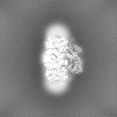
| Images |  emd_16318.png emd_16318.png | 176.9 KB | ||
| Filedesc metadata | emd-16318.cif.gz | 6.7 KB | ||
| Others | emd_16318_half_map_1.map.gzemd_16318_half_map_2.map.gz | 49 MB 49 MB | ||
| Archive directory |  http://ftp.pdbj.org/pub/emdb/structures/EMD-16318ftp://ftp.pdbj.org/pub/emdb/structures/EMD-16318 http://ftp.pdbj.org/pub/emdb/structures/EMD-16318ftp://ftp.pdbj.org/pub/emdb/structures/EMD-16318 | HTTPS FTP |
-Related structure data
| Related structure data | 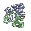 8bxgMC 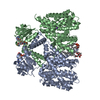 8by2C M: atomic model generated by this map C: citing same article ( |
|---|---|
| Similar structure data |

-Links
| EMDB pages | EMDB (EBI/PDBe) / EMDataResource |
|---|---|
| Related items in Molecule of the Month |

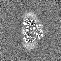
-Map
| File | Download / File: emd_16318.map.gz / Format: CCP4 / Size: 52.7 MB / Type: IMAGE STORED AS FLOATING POINT NUMBER (4 BYTES) | ||||||||||||||||||||||||||||||||||||
|---|---|---|---|---|---|---|---|---|---|---|---|---|---|---|---|---|---|---|---|---|---|---|---|---|---|---|---|---|---|---|---|---|---|---|---|---|---|
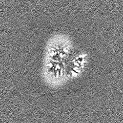
| Projections & slices | Image control
Images are generated by Spider. | ||||||||||||||||||||||||||||||||||||
| Voxel size | X=Y=Z: 0.99675 Å | ||||||||||||||||||||||||||||||||||||
| Density |
| ||||||||||||||||||||||||||||||||||||
| Symmetry | Space group: 1 | ||||||||||||||||||||||||||||||||||||
| Details | EMDB XML:
|
 X (Sec.)
 Y (Row.)
 Z (Col.)


-Supplemental data
-Half map: #2
| File | emd_16318_half_map_1.map | ||||||||||||
|---|---|---|---|---|---|---|---|---|---|---|---|---|---|
| Projections & Slices |
| ||||||||||||
| Density Histograms |


-Half map: #1
| File | emd_16318_half_map_2.map | ||||||||||||
|---|---|---|---|---|---|---|---|---|---|---|---|---|---|
| Projections & Slices |
| ||||||||||||
| Density Histograms |


- Sample components
-Entire : Kefc protein dimer
| Entire | Name: Kefc protein dimer |
|---|---|
| Components |
|
-Supramolecule #1: Kefc protein dimer
| Supramolecule | Name: Kefc protein dimer / type: complex / ID: 1 / Parent: 0 / Macromolecule list: #1 |
|---|---|
| Source (natural) | Organism: Escherichia coli (E. coli) |
-Macromolecule #1: Glutathione-regulated potassium-efflux system protein KefC
| Macromolecule | Name: Glutathione-regulated potassium-efflux system protein KefC type: protein_or_peptide / ID: 1 / Number of copies: 2 / Enantiomer: LEVO |
|---|---|
| Source (natural) | Organism: Escherichia coli (E. coli) |
| Molecular weight | Theoretical: 61.183223 KDa |
| Recombinant expression | Organism: Escherichia coli (E. coli) |
| Sequence | String: MDSHTLIQAL IYLGSAALIV PIAVRLGLGS VLGYLIAGCI IGPWGLRLVT DAESILHFAE IGVVLMLFII GLELDPQRLW KLRAAVFGG GALQMVICGG LLGLFCMLLG LRWQVAELIG MTLALSSTAI AMQAMNERNL MVTQMGRSAF AVLLFQDIAA I PLVAMIPL ...String: MDSHTLIQAL IYLGSAALIV PIAVRLGLGS VLGYLIAGCI IGPWGLRLVT DAESILHFAE IGVVLMLFII GLELDPQRLW KLRAAVFGG GALQMVICGG LLGLFCMLLG LRWQVAELIG MTLALSSTAI AMQAMNERNL MVTQMGRSAF AVLLFQDIAA I PLVAMIPL LATSSASTTM GAFALSALKV AGALVLVVLL GRYVTRPALR FVARSGLREV FSAVALFLVF GFGLLLEEVG LS MAMGAFL AGVLLASSEY RHALESDIEP FKGLLLGLFF IGVGMSIDFG TLLENPLRIV ILLLGFLIIK IAMLWLIARP LQV PNKQRR WFAVLLGQGS EFAFVVFGAA QMANVLEPEW AKSLTLAVAL SMAATPILLV ILNRLEQSST EEAREADEID EEQP RVIIA GFGRFGQITG RLLLSSGVKM VVLDHDPDHI ETLRKFGMKV FYGDATRMDL LESAGAAKAE VLINAIDDPQ TNLQL TEMV KEHFPHLQII ARARDVDHYI RLRQAGVEKP ERETFEGALK TGRLALESLG LGPYEARERA DVFRRFNIQM VEEMAM V UniProtKB: Glutathione-regulated potassium-efflux system protein KefC |
-Macromolecule #2: POTASSIUM ION
| Macromolecule | Name: POTASSIUM ION / type: ligand / ID: 2 / Number of copies: 2 / Formula: K |
|---|---|
| Molecular weight | Theoretical: 39.098 Da |
-Macromolecule #3: ADENOSINE MONOPHOSPHATE
| Macromolecule | Name: ADENOSINE MONOPHOSPHATE / type: ligand / ID: 3 / Number of copies: 2 / Formula: AMP |
|---|---|
| Molecular weight | Theoretical: 347.221 Da |
| Chemical component information |  ChemComp-AMP: |
-Macromolecule #4: (1R)-2-{[(S)-{[(2S)-2,3-dihydroxypropyl]oxy}(hydroxy)phosphoryl]o...
| Macromolecule | Name: (1R)-2-{[(S)-{[(2S)-2,3-dihydroxypropyl]oxy}(hydroxy)phosphoryl]oxy}-1-[(hexadecanoyloxy)methyl]ethyl (9Z)-octadec-9-enoate type: ligand / ID: 4 / Number of copies: 2 / Formula: PGW |
|---|---|
| Molecular weight | Theoretical: 749.007 Da |
-Experimental details
-Structure determination
| Method | cryo EM |
|---|---|
 Processing Processing | single particle reconstruction |
| Aggregation state | particle |
-Sample preparation
| Buffer | pH: 7.5 |
|---|---|
| Grid | Material: COPPER |
| Vitrification | Cryogen name: ETHANE / Chamber humidity: 100 % / Instrument: FEI VITROBOT MARK IV |
- Electron microscopy
| Microscope | FEI TITAN KRIOS |
|---|---|
| Electron beam | Acceleration voltage: 300 kV / Electron source: FIELD EMISSION GUN |
| Electron optics | Illumination mode: FLOOD BEAM / Imaging mode: BRIGHT FIELDBright-field microscopy / Nominal defocus max: 2.0 µm / Nominal defocus min: 0.6 µm |
| Image recording | Film or detector model: GATAN K3 BIOQUANTUM (6k x 4k) / Average electron dose: 52.2 e/Å2 |
| Experimental equipment |  Model: Titan Krios / Image courtesy: FEI Company |
-Image processing
| Startup model | Type of model: NONE |
|---|---|
| Initial angle assignment | Type: MAXIMUM LIKELIHOOD |
| Final angle assignment | Type: MAXIMUM LIKELIHOOD |
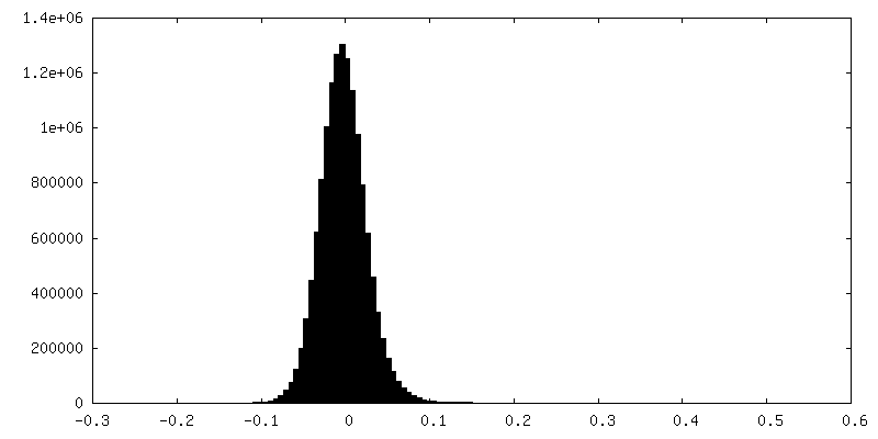
| Final reconstruction | Resolution.type: BY AUTHOR / Resolution: 3.16 Å / Resolution method: FSC 0.143 CUT-OFF / Number images used: 305737 |
| FSC plot (resolution estimation) |  |
