 Movie
Movie Controller

[English] 日本語
 Yorodumi
- EMDB-15860: Cryo-EM structure of ribosome-Sec61-TRAP (TRanslocon Associated P... -
+ Open data

- Basic information
| Entry | 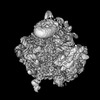 | |||||||||
|---|---|---|---|---|---|---|---|---|---|---|
| Title | Cryo-EM structure of ribosome-Sec61-TRAP (TRanslocon Associated Protein) translocon complex | |||||||||
 Map data Map data | ||||||||||
 Sample Sample |
| |||||||||
| Function / homology |  Function and homology information Function and homology informationcytosolic ribosome /  5S rRNA binding / cytosolic large ribosomal subunit / ribosome / structural constituent of ribosome / ribonucleoprotein complex / translation / nucleolus / RNA binding / cytoplasm 5S rRNA binding / cytosolic large ribosomal subunit / ribosome / structural constituent of ribosome / ribonucleoprotein complex / translation / nucleolus / RNA binding / cytoplasmSimilarity search - Function | |||||||||
| Biological species |  Canis lupus familiaris (dog) / rabbit (rabbit) / Rabbit (rabbit) / Oryctolagus cuniculus (rabbit) Canis lupus familiaris (dog) / rabbit (rabbit) / Rabbit (rabbit) / Oryctolagus cuniculus (rabbit) | |||||||||
| Method | single particle reconstruction / cryo EM / Resolution: 2.86 Å | |||||||||
 Authors Authors | Pauwels E / Shewakramani NR / De Wijngaert B / Vermeire K / Das K | |||||||||
| Funding support |  Belgium, 1 items Belgium, 1 items
| |||||||||
 Citation Citation | Journal: Sci Adv / Year: 2023 Title: Structural insights into TRAP association with ribosome-Sec61 complex and translocon inhibition by a CADA derivative. Authors: Eva Pauwels / Neesha R Shewakramani / Brent De Wijngaert / Anita Camps / Becky Provinciael / Joren Stroobants / Kai-Uwe Kalies / Enno Hartmann / Piet Maes / Kurt Vermeire / Kalyan Das /  Abstract: During cotranslational translocation, the signal peptide of a nascent chain binds Sec61 translocon to initiate protein transport through the endoplasmic reticulum (ER) membrane. Our cryo-electron ...During cotranslational translocation, the signal peptide of a nascent chain binds Sec61 translocon to initiate protein transport through the endoplasmic reticulum (ER) membrane. Our cryo-electron microscopy structure of ribosome-Sec61 shows binding of an ordered heterotetrameric translocon-associated protein (TRAP) complex, in which TRAP-γ is anchored at two adjacent positions of 28 ribosomal RNA and interacts with ribosomal protein L38 and Sec61α/γ. Four transmembrane helices (TMHs) of TRAP-γ cluster with one C-terminal helix of each α, β, and δ subunits. The seven TMH bundle helps position a crescent-shaped trimeric TRAP-α/β/δ core in the ER lumen, facing the Sec61 channel. Further, our in vitro assay establishes the cyclotriazadisulfonamide derivative CK147 as a translocon inhibitor. A structure of ribosome-Sec61-CK147 reveals CK147 binding the channel and interacting with the plug helix from the lumenal side. The CK147 resistance mutations surround the inhibitor. These structures help in understanding the TRAP functions and provide a new Sec61 site for designing translocon inhibitors. | |||||||||
| History |
|
- Structure visualization
| Supplemental images |
|---|

- Downloads & links
-EMDB archive
| Map data | emd_15860.map.gz | 96.4 MB | EMDB map data format | |
|---|---|---|---|---|
| Header (meta data) | emd-15860-v30.xmlemd-15860.xml | 68.2 KB 68.2 KB | Display Display | EMDB header |
| FSC (resolution estimation) | emd_15860_fsc.xml | 12.4 KB | Display | FSC data file |
| Images | 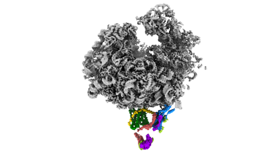 emd_15860.png emd_15860.png | 48.3 KB | ||
| Others | emd_15860_half_map_1.map.gzemd_15860_half_map_2.map.gz | 131.7 MB 131.7 MB | ||
| Archive directory |  http://ftp.pdbj.org/pub/emdb/structures/EMD-15860ftp://ftp.pdbj.org/pub/emdb/structures/EMD-15860 http://ftp.pdbj.org/pub/emdb/structures/EMD-15860ftp://ftp.pdbj.org/pub/emdb/structures/EMD-15860 | HTTPS FTP |
-Related structure data
| Related structure data |  8b5lMC  8b6cC M: atomic model generated by this map C: citing same article ( |
|---|---|
| Similar structure data |

-Links
| EMDB pages | EMDB (EBI/PDBe) / EMDataResource |
|---|---|
| Related items in Molecule of the Month |


-Map
| File | Download / File: emd_15860.map.gz / Format: CCP4 / Size: 166.4 MB / Type: IMAGE STORED AS FLOATING POINT NUMBER (4 BYTES) | ||||||||||||||||||||||||||||||||||||
|---|---|---|---|---|---|---|---|---|---|---|---|---|---|---|---|---|---|---|---|---|---|---|---|---|---|---|---|---|---|---|---|---|---|---|---|---|---|
| Projections & slices | Image control
Images are generated by Spider. | ||||||||||||||||||||||||||||||||||||
| Voxel size | X=Y=Z: 1.23 Å | ||||||||||||||||||||||||||||||||||||
| Density |
| ||||||||||||||||||||||||||||||||||||
| Symmetry | Space group: 1 | ||||||||||||||||||||||||||||||||||||
| Details | EMDB XML:
|
 Z (Sec.)
 Y (Row.)
 X (Col.)


-Supplemental data
-Half map: #2
| File | emd_15860_half_map_1.map | ||||||||||||
|---|---|---|---|---|---|---|---|---|---|---|---|---|---|
| Projections & Slices |
| ||||||||||||
| Density Histograms |


-Half map: #1
| File | emd_15860_half_map_2.map | ||||||||||||
|---|---|---|---|---|---|---|---|---|---|---|---|---|---|
| Projections & Slices |
| ||||||||||||
| Density Histograms |


- Sample components
+Entire : 80S ribosome complex with Sec61 and TRAP
+Supramolecule #1: 80S ribosome complex with Sec61 and TRAP
+Macromolecule #1: 60S ribosomal protein L35
+Macromolecule #2: 60S ribosomal protein L17
+Macromolecule #3: 60S ribosomal protein L31
+Macromolecule #5: 60S ribosomal protein L7a
+Macromolecule #6: Ribosomal protein L32
+Macromolecule #7: Signal sequence receptor subunit 3
+Macromolecule #8: 60S ribosomal protein L18a
+Macromolecule #9: Ribosomal protein L10
+Macromolecule #10: Ribosomal_L18_c domain-containing protein
+Macromolecule #11: 60S ribosomal protein L23
+Macromolecule #12: 60S ribosomal protein L6
+Macromolecule #13: 60S ribosomal protein L11
+Macromolecule #14: Translocon-associated protein subunit alpha
+Macromolecule #15: 60S ribosomal protein L38
+Macromolecule #16: 60S ribosomal protein L29
+Macromolecule #17: Ubiquitin-60S ribosomal protein L40
+Macromolecule #18: 60S ribosomal protein L27
+Macromolecule #19: 60S ribosomal protein L21
+Macromolecule #20: 60S ribosomal protein L36
+Macromolecule #21: 60S ribosomal protein L7
+Macromolecule #22: 60S ribosomal protein L41
+Macromolecule #23: 60S ribosomal protein L13
+Macromolecule #24: 60S ribosomal protein L13a
+Macromolecule #26: 60S ribosomal protein L4
+Macromolecule #27: Ribosomal protein L26
+Macromolecule #28: Ribosomal protein L3
+Macromolecule #29: Ribosomal protein L19
+Macromolecule #30: Ribosomal_L23eN domain-containing protein
+Macromolecule #31: 60S ribosomal protein L37a
+Macromolecule #32: 60S ribosomal protein L14
+Macromolecule #33: 60S ribosomal protein L9
+Macromolecule #35: 60S ribosomal protein L28
+Macromolecule #36: Ribosomal protein L18
+Macromolecule #37: 60S ribosomal protein L35a
+Macromolecule #38: 60S ribosomal protein L27a
+Macromolecule #39: Ribosomal protein L8
+Macromolecule #40: 60S ribosomal protein L34
+Macromolecule #41: Translocon-associated protein subunit delta
+Macromolecule #42: Protein transport protein Sec61 subunit gamma
+Macromolecule #43: Translocon-associated protein subunit beta
+Macromolecule #44: Ribosomal protein L37
+Macromolecule #45: Ribosomal protein L15
+Macromolecule #46: 60S ribosomal protein L36a-like
+Macromolecule #47: Protein transport protein Sec61 subunit alpha isoform 1
+Macromolecule #48: 60S ribosomal protein L30
+Macromolecule #49: Ribosomal protein L24
+Macromolecule #50: 60S ribosomal protein L39-like
+Macromolecule #51: 60S ribosomal protein L22
+Macromolecule #4: 28S rRNA
+Macromolecule #25: 5.8S rRNA
+Macromolecule #34: 5S rRNA
+Macromolecule #52: MAGNESIUM ION
+Macromolecule #53: ZINC ION
-Experimental details
-Structure determination
| Method | cryo EM |
|---|---|
 Processing Processing | single particle reconstruction |
| Aggregation state | particle |
-Sample preparation
| Buffer | pH: 7.5 |
|---|---|
| Grid | Model: Quantifoil R1.2/1.3 / Material: COPPER / Mesh: 300 / Support film - Material: CARBON / Support film - topology: HOLEY / Support film - Film thickness: 2.0 nm / Pretreatment - Type: GLOW DISCHARGE / Pretreatment - Time: 30 sec. / Pretreatment - Atmosphere: AIR / Pretreatment - Pressure: 0.015 kPa |
| Vitrification | Cryogen name: ETHANE / Chamber humidity: 95 % / Chamber temperature: 277.15 K / Instrument: FEI VITROBOT MARK IV |
- Electron microscopy
| Microscope | TFS GLACIOS |
|---|---|
| Electron beam | Acceleration voltage: 200 kV / Electron source: FIELD EMISSION GUN |
| Electron optics | C2 aperture diameter: 50.0 µm / Calibrated magnification: 120000 / Illumination mode: FLOOD BEAM / Imaging mode: BRIGHT FIELDBright-field microscopy / Cs: 2.7 mm / Nominal defocus max: 1.8 µm / Nominal defocus min: 0.6 µm |
| Sample stage | Specimen holder model: OTHER / Cooling holder cryogen: NITROGEN |
| Image recording | Film or detector model: FEI FALCON III (4k x 4k) / Detector mode: COUNTING / Number grids imaged: 1 / Number real images: 2805 / Average exposure time: 40.0 sec. / Average electron dose: 0.8 e/Å2 |
-Image processing
| Particle selection | Number selected: 385028 |
|---|---|
| Startup model | Type of model: PDB ENTRY PDB model - PDB ID: |
| Initial angle assignment | Type: MAXIMUM LIKELIHOOD / Software - Name: RELION (ver. 3.1) |
| Final 3D classification | Software - Name: RELION (ver. 3.1) |
| Final angle assignment | Type: MAXIMUM LIKELIHOOD / Software - Name: RELION (ver. 3.1) |
| Final reconstruction | Applied symmetry - Point group: C1 (asymmetric) / Resolution.type: BY AUTHOR / Resolution: 2.86 Å / Resolution method: FSC 0.143 CUT-OFF / Software - Name: RELION (ver. 3.1) / Number images used: 119208 |
| FSC plot (resolution estimation) |  |


