 Movie
Movie Controller

[English] 日本語
 Yorodumi
- EMDB-15399: Cryo-EM map of crescentin filament in complex with a megabody (st... -
+ Open data

- Basic information
| Entry |  | |||||||||
|---|---|---|---|---|---|---|---|---|---|---|
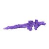
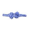
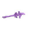
| Title | Cryo-EM map of crescentin filament in complex with a megabody (stutter mutant, C2 symmetry, large box) | |||||||||
 Map data Map data | ||||||||||
 Sample Sample |
| |||||||||
 Keywords Keywords |  cytoskeleton / cell shape / intermediate filaments / coiled coil / assembly / STRUCTURAL PROTEIN cytoskeleton / cell shape / intermediate filaments / coiled coil / assembly / STRUCTURAL PROTEIN | |||||||||
| Biological species |  Caulobacter vibrioides (bacteria) / Caulobacter vibrioides (bacteria) /  Camelidae (mammal) Camelidae (mammal) | |||||||||
| Method | single particle reconstruction / cryo EM / Resolution: 4.1 Å | |||||||||
 Authors Authors | Liu Y / Lowe J | |||||||||
| Funding support |  United Kingdom, 1 items United Kingdom, 1 items
| |||||||||
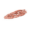
 Citation Citation | Journal: Proc Natl Acad Sci U S A / Year: 2024 Title: Filament structure and subcellular organization of the bacterial intermediate filament-like protein crescentin. Authors: Yue Liu / Fusinita van den Ent / Jan Löwe / Abstract: The protein crescentin is required for the crescent shape of the freshwater bacterium (). Crescentin forms a filamentous structure on the inner, concave side of the curved cells. It shares features ...The protein crescentin is required for the crescent shape of the freshwater bacterium (). Crescentin forms a filamentous structure on the inner, concave side of the curved cells. It shares features with eukaryotic intermediate filament (IF) proteins, including the formation of static filaments based on long and parallel coiled coils, the protein's length, structural roles in cell and organelle shape determination and the presence of a coiled coil discontinuity called the "stutter." Here, we have used electron cryomicroscopy (cryo-EM) to determine the structure of the full-length protein and its filament, exploiting a crescentin-specific nanobody. The filament is formed by two strands, related by twofold symmetry, that each consist of two dimers, resulting in an octameric assembly. Crescentin subunits form longitudinal contacts head-to-head and tail-to-tail, making the entire filament non-polar. Using in vivo site-directed cysteine cross-linking, we demonstrated that contacts observed in the in vitro filament structure exist in cells. Electron cryotomography (cryo-ET) of cells expressing crescentin showed filaments on the concave side of the curved cells, close to the inner membrane, where they form a band. When comparing with current models of IF proteins and their filaments, which are also built from parallel coiled coil dimers and lack overall polarity, it emerges that IF proteins form head-to-tail longitudinal contacts in contrast to crescentin and hence several inter-dimer contacts in IFs have no equivalents in crescentin filaments. Our work supports the idea that intermediate filament-like proteins achieve their shared polymerization and mechanical properties through a variety of filament architectures. #1: Journal: to be publishedTitle: Assembly and organization of the bacterial intermediate filament-like protein crescentin Authors: Liu Y / Lowe J | |||||||||
| History |
|
- Structure visualization
| Supplemental images |
|---|

- Downloads & links
-EMDB archive
| Map data | emd_15399.map.gz | 735.8 MB |  EMDB map data format EMDB map data format | |
|---|---|---|---|---|
| Header (meta data) | emd-15399-v30.xmlemd-15399.xml | 18.5 KB 18.5 KB | Display Display | EMDB header |
| FSC (resolution estimation) | emd_15399_fsc.xml | 20.9 KB | Display | FSC data file |
| Images |  emd_15399.png emd_15399.png | 31.4 KB | ||
| Masks | emd_15399_msk_1.map | 824 MB | Mask map | |
| Filedesc metadata | emd-15399.cif.gz | 5.8 KB | ||
| Others | emd_15399_half_map_1.map.gzemd_15399_half_map_2.map.gz | 765.8 MB 765.8 MB | ||
| Archive directory |  http://ftp.pdbj.org/pub/emdb/structures/EMD-15399ftp://ftp.pdbj.org/pub/emdb/structures/EMD-15399 http://ftp.pdbj.org/pub/emdb/structures/EMD-15399ftp://ftp.pdbj.org/pub/emdb/structures/EMD-15399 | HTTPS FTP |
-Related structure data


-Links
| EMDB pages | EMDB (EBI/PDBe) / EMDataResource |
|---|
-Map
| File | Download / File: emd_15399.map.gz / Format: CCP4 / Size: 824 MB / Type: IMAGE STORED AS FLOATING POINT NUMBER (4 BYTES) | ||||||||||||||||||||
|---|---|---|---|---|---|---|---|---|---|---|---|---|---|---|---|---|---|---|---|---|---|
| Voxel size | X=Y=Z: 1.59 Å | ||||||||||||||||||||
| Density |
| ||||||||||||||||||||
| Symmetry | Space group: 1 | ||||||||||||||||||||
| Details | EMDB XML:
|
-Supplemental data
-Mask #1
| File | emd_15399_msk_1.map | ||||||||||||
|---|---|---|---|---|---|---|---|---|---|---|---|---|---|
| Projections & Slices |
| ||||||||||||
| Density Histograms |
 Z
Z Y
Y X
X


-Half map: #1
| File | emd_15399_half_map_1.map | ||||||||||||
|---|---|---|---|---|---|---|---|---|---|---|---|---|---|
| Projections & Slices |
| ||||||||||||
| Density Histograms |


-Half map: #2
| File | emd_15399_half_map_2.map | ||||||||||||
|---|---|---|---|---|---|---|---|---|---|---|---|---|---|
| Projections & Slices |
| ||||||||||||
| Density Histograms |


- Sample components
-Entire : Crescentin filament in complex with megabody MB13
| Entire | Name: Crescentin filament in complex with megabody MB13 |
|---|---|
| Components |
|
-Supramolecule #1: Crescentin filament in complex with megabody MB13
| Supramolecule | Name: Crescentin filament in complex with megabody MB13 / type: complex / ID: 1 / Parent: 0 / Macromolecule list: all |
|---|
-Supramolecule #2: Caulobacter crescentus strain NA1000 crescentin (stutter mutant)
| Supramolecule | Name: Caulobacter crescentus strain NA1000 crescentin (stutter mutant) type: complex / ID: 2 / Parent: 1 |
|---|---|
| Source (natural) | Organism: Caulobacter vibrioides (bacteria) |
-Supramolecule #3: Crescentin-specific megabody MB13
| Supramolecule | Name: Crescentin-specific megabody MB13 / type: complex / ID: 3 / Parent: 1 |
|---|---|
| Source (natural) | Organism: Camelidae (mammal) |
-Macromolecule #1: Caulobacter crescentus strain NA1000 crescentin (stutter mutant)
| Macromolecule | Name: Caulobacter crescentus strain NA1000 crescentin (stutter mutant) type: protein_or_peptide / ID: 1 / Enantiomer: LEVO |
|---|---|
| Source (natural) | Organism: Caulobacter vibrioides (bacteria) |
| Recombinant expression | Organism: Escherichia coli (E. coli) |
| Sequence | String: MRLLSKNSRE TKNGKPTVLG DEARAEAMQH QIESTQAIGQ RYETIHGGLD SIGRVMEHLK AIEPLIAEIR GPVSQEFEAR RAEHAELIAV RANLDQAQRQ IALIQAEERE VSARLAAAET ALGESDARRQ TQDAALEDNA LEIDRLRNAL LQSDLKVSSL DASLRDATAR ...String: MRLLSKNSRE TKNGKPTVLG DEARAEAMQH QIESTQAIGQ RYETIHGGLD SIGRVMEHLK AIEPLIAEIR GPVSQEFEAR RAEHAELIAV RANLDQAQRQ IALIQAEERE VSARLAAAET ALGESDARRQ TQDAALEDNA LEIDRLRNAL LQSDLKVSSL DASLRDATAR IEHLVQDVEG LRVQAQDIDA RRGDAEAALA RANQDNALLG EEAATLKKRV DQAGLDLARL SRIETDLEAQ LAAERARVQA VENALAAHQA DSGRTIRGLE SQVEANRAEI SALQTRLETA TGRADKLEEM NGQISARLAD SSAQQKAVER RAGDLNVALE RALDRIRALE EEADGLRQRH AGVDTARATA IERADQLAKS AVAQEKALKR AEERAQQLRA RLDAMQEAQD QVRRDSATHE AKIAELQATI ERLTSEAALA EGALEAARRD RSRLQMALLG ASDGDVAASA |
-Macromolecule #2: Crescentin-specific megabody MB13
| Macromolecule | Name: Crescentin-specific megabody MB13 / type: protein_or_peptide / ID: 2 / Enantiomer: LEVO |
|---|---|
| Source (natural) | Organism: Camelidae (mammal) |
| Recombinant expression | Organism: Escherichia coli (E. coli) |
| Sequence | String: EVQLQESGGG LVYKEETQSG LNNYARVVEK GQYDSLEIPA QVAASWESGR DDAAVFGFID KEQLDKYVAN GGKRSDWTVK FAENRSQDGT LLGYSLLQES VDQASYMYSD NHYLAEMATI LGKPEEAKRY RQLAQQLADY INTCMFDPTT QFYYDVRIED KPLANGCAGK ...String: EVQLQESGGG LVYKEETQSG LNNYARVVEK GQYDSLEIPA QVAASWESGR DDAAVFGFID KEQLDKYVAN GGKRSDWTVK FAENRSQDGT LLGYSLLQES VDQASYMYSD NHYLAEMATI LGKPEEAKRY RQLAQQLADY INTCMFDPTT QFYYDVRIED KPLANGCAGK PIVERGKGPE GWSPLFNGAA TQANADAVVK VMLDPKEFNT FVPLGTAALT NPAFGADIYW RGRVWVDQFW FGLKGMERYG YRDDALKLAD TFFRHAKGLT ADGPIQENYN PLTGAQQGAP NFSWSAAHLY MLYNDFFRKQ ASGGGSGGGG SGGGGSGNAD NYKNVINRTG APQYMKDYDY DDHQRFNPFF DLGAWHGHLL PDGPNTMGGF PGVALLTEEY INFMASNFDR LTVWQDGKKV DFTLEAYSIP GALVQKLTAK DVQVEMTLRF ATPRTSLLET KITSNKPLDL VWDGELLEKL EAKEGKPLSD KTIAGEYPDY QRKISATRDG LKVTFGKVRA TWDLLTSGES EYQVHKSLPV QTEINGNRFT SKAHINGSTT LYTTYSHLLT AQEVSKEQMQ IRDILARPAF YLTASQQRWE EYLKKGLTNP DATPEQTRVA VKAIETLNGN WRSPGGAVKF NTVTPSVTGR WFSGNQTWPW DTWKQAFAMA HFNPDIAKEN IRAVFSWQIQ PGDSVRPQDV GFVPDLIAWN LSPERGGDGG NWNERNTKPS LAAWSVMEVY NVTQDKTWVA EMYPKLVAYH DWWLRNRDHN GNGVPEYGAT RDKAHNTESG EMLFTVKKDS LRLSCASSRS IDGINIMRWY RQAPGKQRGM VAVVTGWGST NYVDSVKGRF IISRDSAKDT VYLQMNNLKP EDTAVYSCNA IYRGSEYWGQ GTQVTVSSGE NLYFQGSHHH HHHHHHH |
-Experimental details
-Structure determination
| Method | cryo EM |
|---|---|
 Processing Processing | single particle reconstruction |
| Aggregation state | filament |
-Sample preparation
| Concentration | 2 mg/mL |
|---|---|
| Buffer | pH: 6.5 / Details: PIPES 25mM pH 6.5, 0.05% CHAPS |
| Grid | Model: UltrAuFoil R2/2 / Material: GOLD / Mesh: 300 |
| Vitrification | Cryogen name: ETHANE / Chamber humidity: 100 % / Chamber temperature: 283 K / Instrument: FEI VITROBOT MARK IV |
- Electron microscopy
| Microscope | FEI TITAN KRIOS |
|---|---|
| Electron beam | Acceleration voltage: 300 kV / Electron source: FIELD EMISSION GUN |
| Electron optics | C2 aperture diameter: 70.0 µm / Illumination mode: FLOOD BEAM / Imaging mode: BRIGHT FIELDBright-field microscopy / Cs: 2.7 mm / Nominal defocus max: 2.4 µm / Nominal defocus min: 0.8 µm / Nominal magnification: 81000 |
| Specialist optics | Energy filter - Name: GIF Bioquantum / Energy filter - Slit width: 20 eV |
| Sample stage | Specimen holder model: FEI TITAN KRIOS AUTOGRID HOLDER / Cooling holder cryogen: NITROGEN |
| Temperature | Min: 80.0 K / Max: 80.0 K |
| Image recording | Film or detector model: GATAN K3 (6k x 4k) / Number grids imaged: 2 / Average exposure time: 2.4 sec. / Average electron dose: 53.0 e/Å2 |
| Experimental equipment |  Model: Titan Krios / Image courtesy: FEI Company |
-Image processing
| Startup model | Type of model: NONE |
|---|---|
| Initial angle assignment | Type: RANDOM ASSIGNMENT / Software - Name: cryoSPARC |
| Final 3D classification | Software - Name: cryoSPARC |
| Final angle assignment | Type: PROJECTION MATCHING / Software: (Name: cryoSPARC, RELION) |
| Final reconstruction | Applied symmetry - Point group: C2 (2 fold cyclic) / Algorithm: FOURIER SPACE / Resolution.type: BY AUTHOR / Resolution: 4.1 Å / Resolution method: FSC 0.143 CUT-OFF / Software - Name: cryoSPARC / Number images used: 585252 |
| FSC plot (resolution estimation) |  |
