 ムービー
ムービー コントローラー


+ データを開く

- 基本情報
基本情報
| 登録情報 | データベース: EMDB / ID: EMD-1480 | |||||||||
|---|---|---|---|---|---|---|---|---|---|---|
| タイトル | 3D structure of the canine 80S ribosome | |||||||||
 マップデータ マップデータ | 3D volume of the canine 80S ribosome determined at 8.7 A resolution (Fsc 0.5), within the context of a ribosome-channel complex from ER membranes. | |||||||||
 試料 試料 |
| |||||||||
 キーワード キーワード |  eukaryotic ribosome / protein translation (翻訳 (生物学)) / tRNA translocation / expansion segments eukaryotic ribosome / protein translation (翻訳 (生物学)) / tRNA translocation / expansion segments | |||||||||
| 生物種 |  Canis lupus familiaris (イヌ) Canis lupus familiaris (イヌ) | |||||||||
| 手法 | 単粒子再構成法 / クライオ電子顕微鏡法 / 解像度: 8.7 Å | |||||||||
 データ登録者 データ登録者 | Chandramouli P / Topf M / Menetret JF / Eswar N / Cannone JJ / Gutell R / Sali A / Akey CW | |||||||||
 引用 引用 | ジャーナル: Structure / 年: 2008 タイトル: Structure of the mammalian 80S ribosome at 8.7 A resolution. 著者: Preethi Chandramouli / Maya Topf / Jean-François Ménétret / Narayanan Eswar / Jamie J Cannone / Robin R Gutell / Andrej Sali / Christopher W Akey /  要旨: In this paper, we present a structure of the mammalian ribosome determined at approximately 8.7 A resolution by electron cryomicroscopy and single-particle methods. A model of the ribosome was ...In this paper, we present a structure of the mammalian ribosome determined at approximately 8.7 A resolution by electron cryomicroscopy and single-particle methods. A model of the ribosome was created by docking homology models of subunit rRNAs and conserved proteins into the density map. We then modeled expansion segments in the subunit rRNAs and found unclaimed density for approximately 20 proteins. In general, many conserved proteins and novel proteins interact with expansion segments to form an integrated framework that may stabilize the mature ribosome. Our structure provides a snapshot of the mammalian ribosome at the beginning of translation and lends support to current models in which large movements of the small subunit and L1 stalk occur during tRNA translocation. Finally, details are presented for intersubunit bridges that are specific to the eukaryotic ribosome. We suggest that these bridges may help reset the conformation of the ribosome to prepare for the next cycle of chain elongation. | |||||||||
| 履歴 |
|
- 構造の表示
構造の表示
| ムービー |
 ムービービューア ムービービューア |
|---|---|
| 構造ビューア | EMマップ: SurfViewMolmilJmol/JSmol |
| 添付画像 |

- ダウンロードとリンク
-EMDBアーカイブ
| マップデータ | emd_1480.map.gz | 4 MB | EMDBマップデータ形式 | |
|---|---|---|---|---|
| ヘッダ (付随情報) | emd-1480-v30.xmlemd-1480.xml | 10.4 KB 10.4 KB | 表示 表示 | EMDBヘッダ |
| 画像 | 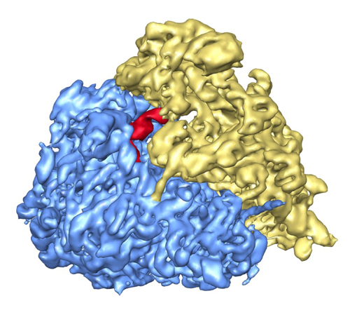 1480.jpg 1480.jpg | 169.3 KB | ||
| アーカイブディレクトリ |  http://ftp.pdbj.org/pub/emdb/structures/EMD-1480ftp://ftp.pdbj.org/pub/emdb/structures/EMD-1480 http://ftp.pdbj.org/pub/emdb/structures/EMD-1480ftp://ftp.pdbj.org/pub/emdb/structures/EMD-1480 | HTTPS FTP |
-関連構造データ


-リンク
| EMDBのページ | EMDB (EBI/PDBe) / EMDataResource |
|---|---|
| 「今月の分子」の関連する項目 |

-マップ
| ファイル | ダウンロード / ファイル: emd_1480.map.gz / 形式: CCP4 / 大きさ: 17.7 MB / タイプ: IMAGE STORED AS FLOATING POINT NUMBER (4 BYTES) | ||||||||||||||||||||||||||||||||||||||||||||||||||||||||||||||||||||
|---|---|---|---|---|---|---|---|---|---|---|---|---|---|---|---|---|---|---|---|---|---|---|---|---|---|---|---|---|---|---|---|---|---|---|---|---|---|---|---|---|---|---|---|---|---|---|---|---|---|---|---|---|---|---|---|---|---|---|---|---|---|---|---|---|---|---|---|---|---|
| 注釈 | 3D volume of the canine 80S ribosome determined at 8.7 A resolution (Fsc 0.5), within the context of a ribosome-channel complex from ER membranes. | ||||||||||||||||||||||||||||||||||||||||||||||||||||||||||||||||||||
| 投影像・断面図 | 画像のコントロール
画像は Spider により作成 | ||||||||||||||||||||||||||||||||||||||||||||||||||||||||||||||||||||
| ボクセルのサイズ | X=Y=Z: 2.73 Å | ||||||||||||||||||||||||||||||||||||||||||||||||||||||||||||||||||||
| 密度 |
| ||||||||||||||||||||||||||||||||||||||||||||||||||||||||||||||||||||
| 対称性 | 空間群: 1 | ||||||||||||||||||||||||||||||||||||||||||||||||||||||||||||||||||||
| 詳細 | EMDB XML:
CCP4マップ ヘッダ情報:
| ||||||||||||||||||||||||||||||||||||||||||||||||||||||||||||||||||||
 Z (Sec.)
 Y (Row.)
 X (Col.)


-添付データ
- 試料の構成要素
-全体 : canine 80S ribosome
| 全体 | 名称: canine 80S ribosome |
|---|---|
| 要素 |
|
-超分子 #1000: canine 80S ribosome
| 超分子 | 名称: canine 80S ribosome / タイプ: sample / ID: 1000 / 詳細: Sample was monodisperse with some mild aggregation. / 集合状態: monomer / Number unique components: 1 |
|---|---|
| 分子量 | 理論値: 3.6 MDa |
-超分子 #1: 80S ribosome
| 超分子 | 名称: 80S ribosome / タイプ: complex / ID: 1 / Name.synonym: ribosome 詳細: The ribosome structure was determined within a larger, ribosome-channel complex isolated from ER membranes. 組換発現: No / Ribosome-details: ribosome-eukaryote: ALL |
|---|---|
| 由来(天然) | 生物種: Canis lupus familiaris (イヌ) / 別称: Dog |
| 分子量 | 理論値: 3.6 MDa |
-実験情報
-構造解析
| 手法 | クライオ電子顕微鏡法 |
|---|---|
 解析 解析 | 単粒子再構成法 |
| 試料の集合状態 | particle |
-試料調製
| 緩衝液 | pH: 7.5 詳細: 30mM Hepesm 50mM KAc, 10mM Mg acetate and 1.5% digitonin. |
|---|---|
| グリッド | 詳細: 400 |
| 凍結 | 凍結剤: ETHANE / チャンバー内湿度: 90 % / 装置: HOMEMADE PLUNGER 詳細: Vitrification instrument: home made plunger. in cold room 手法: blot for 1 second |
- 電子顕微鏡法
| 顕微鏡 | FEI TECNAI 20 |
|---|---|
| 電子線 | 加速電圧: 200 kV / 電子線源: FIELD EMISSION GUN |
| 電子光学系 | 倍率(補正後): 51000 / 照射モード: FLOOD BEAM / 撮影モード: BRIGHT FIELDBright-field microscopy / Cs: 2 mm / 最大 デフォーカス(公称値): 3.0 µm / 最小 デフォーカス(公称値): 0.5 µm / 倍率(公称値): 50000 |
| 試料ステージ | 試料ホルダー: eucentric / 試料ホルダーモデル: OTHER |
| 温度 | 平均: 93 K |
| 詳細 | data were collected on Oxford and a Gatan cryo-holders |
| 撮影 | カテゴリ: FILM / フィルム・検出器のモデル: KODAK SO-163 FILM / デジタル化 - スキャナー: OTHER / デジタル化 - サンプリング間隔: 4.5 µm / 実像数: 500 / 平均電子線量: 20 e/Å2 / 詳細: Creoscitex Eversmart was used to scan negatives. / Od range: 1 / ビット/ピクセル: 16 |
-画像解析
| CTF補正 | 詳細: by micrograph |
|---|---|
| 最終 角度割当 | 詳細: 2 degree steps between classes |
| 最終 再構成 | 想定した対称性 - 点群: C1 (非対称) / アルゴリズム: OTHER / 解像度のタイプ: BY AUTHOR / 解像度: 8.7 Å / 解像度の算出法: FSC 0.5 CUT-OFF / ソフトウェア - 名称: EMAN 詳細: sep option equals 3 and setsf were used in the final cycle. a combined structure factor was used to correct for amplitudes in EMAN using the low resolution region from the images and the mid- ...詳細: sep option equals 3 and setsf were used in the final cycle. a combined structure factor was used to correct for amplitudes in EMAN using the low resolution region from the images and the mid-resolution region from a low angle X-ray diffraction pattern. 使用した粒子像数: 78800 |
| 詳細 | Particles selected with Boxer |


