 Movie
Movie Controller


+ Open data

- Basic information
| Entry | Database: EMDB / ID: EMD-1464 | |||||||||
|---|---|---|---|---|---|---|---|---|---|---|
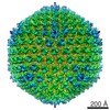
| Title | Adenovirus serotype 5 hexon mediates liver gene transfer. | |||||||||
 Map data Map data | Three-dimensional reconstruction of Adenovirus type 5 bound to Factor X | |||||||||
 Sample Sample |
| |||||||||
| Biological species |    Human adenovirus 5 Human adenovirus 5 | |||||||||
| Method | single particle reconstruction / cryo EM / Resolution: 23.0 Å | |||||||||
 Authors Authors | Waddington SN / McVey JH / Bhella D / Parker AL / Barker K / Atoda H / Pink R / Buckley SM / Greig JA / Denby L ...Waddington SN / McVey JH / Bhella D / Parker AL / Barker K / Atoda H / Pink R / Buckley SM / Greig JA / Denby L / Custers J / Morita T / Francischetti IM / Monteiro RQ / Barouch DH / van Rooijen N / Napoli C / Havenga MJ / Nicklin SA / Baker AH | |||||||||
 Citation Citation | Journal: Cell / Year: 2008 Title: Adenovirus serotype 5 hexon mediates liver gene transfer. Authors: Simon N Waddington / John H McVey / David Bhella / Alan L Parker / Kristeen Barker / Hideko Atoda / Rebecca Pink / Suzanne M K Buckley / Jenny A Greig / Laura Denby / Jerome Custers / ...Authors: Simon N Waddington / John H McVey / David Bhella / Alan L Parker / Kristeen Barker / Hideko Atoda / Rebecca Pink / Suzanne M K Buckley / Jenny A Greig / Laura Denby / Jerome Custers / Takashi Morita / Ivo M B Francischetti / Robson Q Monteiro / Dan H Barouch / Nico van Rooijen / Claudio Napoli / Menzo J E Havenga / Stuart A Nicklin / Andrew H Baker /  Abstract: Adenoviruses are used extensively as gene transfer agents, both experimentally and clinically. However, targeting of liver cells by adenoviruses compromises their potential efficacy. In cell culture, ...Adenoviruses are used extensively as gene transfer agents, both experimentally and clinically. However, targeting of liver cells by adenoviruses compromises their potential efficacy. In cell culture, the adenovirus serotype 5 fiber protein engages the coxsackievirus and adenovirus receptor (CAR) to bind cells. Paradoxically, following intravascular delivery, CAR is not used for liver transduction, implicating alternate pathways. Recently, we demonstrated that coagulation factor (F)X directly binds adenovirus leading to liver infection. Here, we show that FX binds to the Ad5 hexon, not fiber, via an interaction between the FX Gla domain and hypervariable regions of the hexon surface. Binding occurs in multiple human adenovirus serotypes. Liver infection by the FX-Ad5 complex is mediated through a heparin-binding exosite in the FX serine protease domain. This study reveals an unanticipated function for hexon in mediating liver gene transfer in vivo. | |||||||||
| History |
|
- Structure visualization
| Movie |
 Movie viewer Movie viewer |
|---|---|
| Structure viewer | EM map: SurfViewMolmilJmol/JSmol |
| Supplemental images |

- Downloads & links
-EMDB archive
| Map data | emd_1464.map.gz | 20.8 MB | EMDB map data format | |
|---|---|---|---|---|
| Header (meta data) | emd-1464-v30.xmlemd-1464.xml | 10.8 KB 10.8 KB | Display Display | EMDB header |
| Images |  1464.gif 1464.gif | 36.6 KB | ||
| Archive directory |  http://ftp.pdbj.org/pub/emdb/structures/EMD-1464ftp://ftp.pdbj.org/pub/emdb/structures/EMD-1464 http://ftp.pdbj.org/pub/emdb/structures/EMD-1464ftp://ftp.pdbj.org/pub/emdb/structures/EMD-1464 | HTTPS FTP |
-Related structure data
| Similar structure data |
|---|


-Links
| EMDB pages | EMDB (EBI/PDBe) / EMDataResource |
|---|
-Map
| File | Download / File: emd_1464.map.gz / Format: CCP4 / Size: 42.4 MB / Type: IMAGE STORED AS FLOATING POINT NUMBER (4 BYTES) | ||||||||||||||||||||||||||||||||||||||||||||||||||||||||||||||||||||
|---|---|---|---|---|---|---|---|---|---|---|---|---|---|---|---|---|---|---|---|---|---|---|---|---|---|---|---|---|---|---|---|---|---|---|---|---|---|---|---|---|---|---|---|---|---|---|---|---|---|---|---|---|---|---|---|---|---|---|---|---|---|---|---|---|---|---|---|---|---|
| Annotation | Three-dimensional reconstruction of Adenovirus type 5 bound to Factor X | ||||||||||||||||||||||||||||||||||||||||||||||||||||||||||||||||||||
| Projections & slices | Image control
Images are generated by Spider. | ||||||||||||||||||||||||||||||||||||||||||||||||||||||||||||||||||||
| Voxel size | X=Y=Z: 6.54 Å | ||||||||||||||||||||||||||||||||||||||||||||||||||||||||||||||||||||
| Density |
| ||||||||||||||||||||||||||||||||||||||||||||||||||||||||||||||||||||
| Symmetry | Space group: 1 | ||||||||||||||||||||||||||||||||||||||||||||||||||||||||||||||||||||
| Details | EMDB XML:
CCP4 map header:
| ||||||||||||||||||||||||||||||||||||||||||||||||||||||||||||||||||||
 Z (Sec.)
 Y (Row.)
 X (Col.)


-Supplemental data
- Sample components
-Entire : Adenovirus type 5 bound to Factor X
| Entire | Name: Adenovirus type 5 bound to Factor X |
|---|---|
| Components |
|
-Supramolecule #1000: Adenovirus type 5 bound to Factor X
| Supramolecule | Name: Adenovirus type 5 bound to Factor X / type: sample / ID: 1000 / Number unique components: 2 |
|---|
-Supramolecule #1: Human adenovirus 5
| Supramolecule | Name: Human adenovirus 5 / type: virus / ID: 1 / Name.synonym: Ad5 / NCBI-ID: 28285 / Sci species name: Human adenovirus 5 / Virus type: VIRION / Virus isolate: SEROTYPE / Virus enveloped: No / Virus empty: No / Syn species name: Ad5 |
|---|---|
| Host (natural) | Organism:  Homo sapiens (human) / synonym: VERTEBRATES Homo sapiens (human) / synonym: VERTEBRATES |
| Virus shell | Shell ID: 1 / Name: Ad5 / Diameter: 900 Å / T number (triangulation number): 25 |
-Macromolecule #1: Factor X
| Macromolecule | Name: Factor X / type: ligand / ID: 1 / Name.synonym: FX Details: Commercially purified Factor X from Haematologic Technologies Inc. Number of copies: 240 / Oligomeric state: monomer / Recombinant expression: No |
|---|---|
| Source (natural) | Tissue: blood/liver |
-Experimental details
-Structure determination
| Method | cryo EM |
|---|---|
 Processing Processing | single particle reconstruction |
| Aggregation state | particle |
-Sample preparation
| Buffer | pH: 7.4 / Details: 10 nM Tris 100 mM NaCl 5 mM CaCl |
|---|---|
| Grid | Details: 400 mesh copper R2/2 quantifoils |
| Vitrification | Cryogen name: ETHANE Method: 5 ul of virus-ligand complex was loaded onto a freshly glow-discharged grid and blotted until the filter paper detached. |
- Electron microscopy
| Microscope | JEOL 1200EXII |
|---|---|
| Electron beam | Acceleration voltage: 120 kV / Electron source: LAB6 |
| Electron optics | Calibrated magnification: 29128 / Illumination mode: FLOOD BEAM / Imaging mode: BRIGHT FIELDBright-field microscopy / Cs: 3.4 mm / Nominal defocus max: 3.9 µm / Nominal defocus min: 0.668 µm / Nominal magnification: 30000 |
| Sample stage | Specimen holder: side entry, liquid nitrogen cooled / Specimen holder model: OTHER |
| Temperature | Average: 108 K |
| Alignment procedure | Legacy - Astigmatism: Astigmatism corrected at 100,000 times magnification |
| Details | Defocus Pairs were recorded |
| Date | Jun 1, 2007 |
| Image recording | Category: FILM / Film or detector model: KODAK SO-163 FILM / Digitization - Scanner: OTHER / Digitization - Sampling interval: 6.35 µm / Number real images: 21 / Average electron dose: 10 e/Å2 / Details: Images were binned by a factor of 3 / Bits/pixel: 16 |
-Image processing
| CTF correction | Details: each particle was corrected and defocus paired particles merged |
|---|---|
| Final reconstruction | Applied symmetry - Point group: I (icosahedral) / Algorithm: OTHER / Resolution.type: BY AUTHOR / Resolution: 23.0 Å / Resolution method: OTHER / Software - Name: PFT3DR2, MRC / Number images used: 305 |
