 Movie
Movie Controller


+ Open data

- Basic information
| Entry | Database: EMDB / ID: EMD-14369 | |||||||||
|---|---|---|---|---|---|---|---|---|---|---|
| Title | Cryo-EM structure of USP9X, local refinement of monomer | |||||||||
 Map data Map data | sharpened map | |||||||||
 Sample Sample |
| |||||||||
| Function / homology |  Function and homology information Function and homology information cytosolic ciliogenesis / K11-linked deubiquitinase activity / positive regulation of TORC2 signaling / protein deubiquitination involved in ubiquitin-dependent protein catabolic process / protein import into peroxisome matrix, receptor recycling / co-SMAD binding / female gamete generation / : / monoubiquitinated protein deubiquitination / deubiquitinase activity ...cytosolic ciliogenesis / K11-linked deubiquitinase activity / positive regulation of TORC2 signaling / protein deubiquitination involved in ubiquitin-dependent protein catabolic process / protein import into peroxisome matrix, receptor recycling / co-SMAD binding / female gamete generation / : / monoubiquitinated protein deubiquitination / deubiquitinase activity / molecular sequestering activity / axon extension / protein K63-linked deubiquitination / K48-linked deubiquitinase activity / K63-linked deubiquitinase activity / RHOV GTPase cycle / protein deubiquitination / RHOU GTPase cycle / cilium assembly / negative regulation of proteasomal ubiquitin-dependent protein catabolic process / BMP signaling pathway / cysteine-type peptidase activity / Synthesis of active ubiquitin: roles of E1 and E2 enzymes / transforming growth factor beta receptor signaling pathway / chromosome segregation / Peroxisomal protein import / Downregulation of SMAD2/3:SMAD4 transcriptional activity / neuron migration / protein localization / regulation of circadian rhythm / cilium / rhythmic process / cell migration / positive regulation of protein binding / growth cone / ubiquitinyl hydrolase 1 / cysteine-type deubiquitinase activity / amyloid fibril formation / protein stabilization / protein ubiquitination / Ub-specific processing proteases / Amyloid fiber formation / cell division / cysteine-type endopeptidase activity / centrosome / negative regulation of transcription by RNA polymerase II / membrane / nucleus / cytosol / cytoplasm cytosolic ciliogenesis / K11-linked deubiquitinase activity / positive regulation of TORC2 signaling / protein deubiquitination involved in ubiquitin-dependent protein catabolic process / protein import into peroxisome matrix, receptor recycling / co-SMAD binding / female gamete generation / : / monoubiquitinated protein deubiquitination / deubiquitinase activity ...cytosolic ciliogenesis / K11-linked deubiquitinase activity / positive regulation of TORC2 signaling / protein deubiquitination involved in ubiquitin-dependent protein catabolic process / protein import into peroxisome matrix, receptor recycling / co-SMAD binding / female gamete generation / : / monoubiquitinated protein deubiquitination / deubiquitinase activity / molecular sequestering activity / axon extension / protein K63-linked deubiquitination / K48-linked deubiquitinase activity / K63-linked deubiquitinase activity / RHOV GTPase cycle / protein deubiquitination / RHOU GTPase cycle / cilium assembly / negative regulation of proteasomal ubiquitin-dependent protein catabolic process / BMP signaling pathway / cysteine-type peptidase activity / Synthesis of active ubiquitin: roles of E1 and E2 enzymes / transforming growth factor beta receptor signaling pathway / chromosome segregation / Peroxisomal protein import / Downregulation of SMAD2/3:SMAD4 transcriptional activity / neuron migration / protein localization / regulation of circadian rhythm / cilium / rhythmic process / cell migration / positive regulation of protein binding / growth cone / ubiquitinyl hydrolase 1 / cysteine-type deubiquitinase activity / amyloid fibril formation / protein stabilization / protein ubiquitination / Ub-specific processing proteases / Amyloid fiber formation / cell division / cysteine-type endopeptidase activity / centrosome / negative regulation of transcription by RNA polymerase II / membrane / nucleus / cytosol / cytoplasmSimilarity search - Function | |||||||||
| Biological species |  Homo sapiens (human) Homo sapiens (human) | |||||||||
| Method | single particle reconstruction / cryo EM / Resolution: 3.1 Å | |||||||||
 Authors Authors | Deme JC / Halabelian L / Arrowsmith CH / Lea SM / Structural Genomics Consortium (SGC) | |||||||||
| Funding support |  United States, 1 items United States, 1 items
| |||||||||
 Citation Citation | Journal: To Be Published Title: Cryo-EM structure of USP9X Authors: Halabelian L / Deme JC / Lea SM / Arrowsmith CH / Structural Genomics Consortium (SGC) | |||||||||
| History |
|
- Structure visualization
| Movie |
Movie viewer |
|---|---|
| Structure viewer | EM map: SurfViewMolmilJmol/JSmol |
| Supplemental images |

- Downloads & links
-EMDB archive
| Map data | emd_14369.map.gz | 324.1 MB | EMDB map data format | |
|---|---|---|---|---|
| Header (meta data) | emd-14369-v30.xmlemd-14369.xml | 18 KB 18 KB | Display Display | EMDB header |
| FSC (resolution estimation) | emd_14369_fsc.xml | 15.9 KB | Display | FSC data file |
| Images | 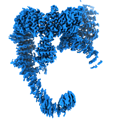 emd_14369.png emd_14369.png | 131.2 KB | ||
| Masks | emd_14369_msk_1.map | 343 MB | Mask map | |
| Others | emd_14369_additional_1.map.gzemd_14369_half_map_1.map.gzemd_14369_half_map_2.map.gz | 171.8 MB 318.2 MB 318.2 MB | ||
| Archive directory |  http://ftp.pdbj.org/pub/emdb/structures/EMD-14369ftp://ftp.pdbj.org/pub/emdb/structures/EMD-14369 http://ftp.pdbj.org/pub/emdb/structures/EMD-14369ftp://ftp.pdbj.org/pub/emdb/structures/EMD-14369 | HTTPS FTP |
-Related structure data
| Related structure data |  7yxyMC  7yxxC C: citing same article ( M: atomic model generated by this map |
|---|---|
| Similar structure data |


-Links
| EMDB pages | EMDB (EBI/PDBe) / EMDataResource |
|---|---|
| Related items in Molecule of the Month |


-Map
| File | Download / File: emd_14369.map.gz / Format: CCP4 / Size: 343 MB / Type: IMAGE STORED AS FLOATING POINT NUMBER (4 BYTES) | ||||||||||||||||||||||||||||||||||||||||||||||||||||||||||||||||||||
|---|---|---|---|---|---|---|---|---|---|---|---|---|---|---|---|---|---|---|---|---|---|---|---|---|---|---|---|---|---|---|---|---|---|---|---|---|---|---|---|---|---|---|---|---|---|---|---|---|---|---|---|---|---|---|---|---|---|---|---|---|---|---|---|---|---|---|---|---|---|
| Annotation | sharpened map | ||||||||||||||||||||||||||||||||||||||||||||||||||||||||||||||||||||
| Projections & slices | Image control
Images are generated by Spider. | ||||||||||||||||||||||||||||||||||||||||||||||||||||||||||||||||||||
| Voxel size | X=Y=Z: 0.832 Å | ||||||||||||||||||||||||||||||||||||||||||||||||||||||||||||||||||||
| Density |
| ||||||||||||||||||||||||||||||||||||||||||||||||||||||||||||||||||||
| Symmetry | Space group: 1 | ||||||||||||||||||||||||||||||||||||||||||||||||||||||||||||||||||||
| Details | EMDB XML:
CCP4 map header:
| ||||||||||||||||||||||||||||||||||||||||||||||||||||||||||||||||||||
 Z (Sec.)
 Y (Row.)
 X (Col.)


-Supplemental data
-Mask #1
| File | emd_14369_msk_1.map | ||||||||||||
|---|---|---|---|---|---|---|---|---|---|---|---|---|---|
| Projections & Slices |
| ||||||||||||
| Density Histograms |


-Additional map: unsharpened map
| File | emd_14369_additional_1.map | ||||||||||||
|---|---|---|---|---|---|---|---|---|---|---|---|---|---|
| Annotation | unsharpened map | ||||||||||||
| Projections & Slices |
| ||||||||||||
| Density Histograms |


-Half map: half map 1
| File | emd_14369_half_map_1.map | ||||||||||||
|---|---|---|---|---|---|---|---|---|---|---|---|---|---|
| Annotation | half map 1 | ||||||||||||
| Projections & Slices |
| ||||||||||||
| Density Histograms |


-Half map: half map 2
| File | emd_14369_half_map_2.map | ||||||||||||
|---|---|---|---|---|---|---|---|---|---|---|---|---|---|
| Annotation | half map 2 | ||||||||||||
| Projections & Slices |
| ||||||||||||
| Density Histograms |


- Sample components
-Entire : USP9X monomer
| Entire | Name: USP9X monomer |
|---|---|
| Components |
|
-Supramolecule #1: USP9X monomer
| Supramolecule | Name: USP9X monomer / type: organelle_or_cellular_component / ID: 1 / Parent: 0 / Macromolecule list: all |
|---|---|
| Source (natural) | Organism: Homo sapiens (human) |
| Recombinant expression | Organism:   Spodoptera frugiperda (fall armyworm) Spodoptera frugiperda (fall armyworm) |
-Macromolecule #1: Probable ubiquitin carboxyl-terminal hydrolase FAF-X
| Macromolecule | Name: Probable ubiquitin carboxyl-terminal hydrolase FAF-X / type: protein_or_peptide / ID: 1 / Number of copies: 1 / Enantiomer: LEVO / EC number: ubiquitinyl hydrolase 1 |
|---|---|
| Source (natural) | Organism: Homo sapiens (human) |
| Molecular weight | Theoretical: 293.878844 KDa |
| Recombinant expression | Organism: Spodoptera frugiperda (fall armyworm) |
| Sequence | String: MHHHHHHSSG RENLYFQGMT ATTRGSPVGG NDNQGQAPDG QSQPPLQQNQ TSSPDSSNEN SPATPPDEQG QGDAPPQLED EEPAFPHTD LAKLDDMINR PRWVVPVLPK GELEVLLEAA IDLSKKGLDV KSEACQRFFR DGLTISFTKI LTDEAVSGWK F EIHRCIIN ...String: MHHHHHHSSG RENLYFQGMT ATTRGSPVGG NDNQGQAPDG QSQPPLQQNQ TSSPDSSNEN SPATPPDEQG QGDAPPQLED EEPAFPHTD LAKLDDMINR PRWVVPVLPK GELEVLLEAA IDLSKKGLDV KSEACQRFFR DGLTISFTKI LTDEAVSGWK F EIHRCIIN NTHRLVELCV AKLSQDWFPL LELLAMALNP HCKFHIYNGT RPCESVSSSV QLPEDELFAR SPDPRSPKGW LV DLLNKFG TLNGFQILHD RFINGSALNV QIIAALIKPF GQCYEFLTLH TVKKYFLPII EMVPQFLENL TDEELKKEAK NEA KNDALS MIIKSLKNLA SRVPGQEETV KNLEIFRLKM ILRLLQISSF NGKMNALNEV NKVISSVSYY THRHGNPEEE EWLT AERMA EWIQQNNILS IVLRDSLHQP QYVEKLEKIL RFVIKEKALT LQDLDNIWAA QAGKHEAIVK NVHDLLAKLA WDFSP EQLD HLFDCFKASW TNASKKQREK LLELIRRLAE DDKDGVMAHK VLNLLWNLAH SDDVPVDIMD LALSAHIKIL DYSCSQ DRD TQKIQWIDRF IEELRTNDKW VIPALKQIRE ICSLFGEAPQ NLSQTQRSPH VFYRHDLINQ LQHNHALVTL VAENLAT YM ESMRLYARDH EDYDPQTVRL GSRYSHVQEV QERLNFLRFL LKDGQLWLCA PQAKQIWKCL AENAVYLCDR EACFKWYS K LMGDEPDLDP DINKDFFESN VLQLDPSLLT ENGMKCFERF FKAVNCREGK LVAKRRAYMM DDLELIGLDY LWRVVIQSN DDIASRAIDL LKEIYTNLGP RLQVNQVVIH EDFIQSCFDR LKASYDTLCV LDGDKDSVNC ARQEAVRMVR VLTVLREYIN ECDSDYHEE RTILPMSRAF RGKHLSFVVR FPNQGRQVDD LEVWSHTNDT IGSVRRCILN RIKANVAHTK IELFVGGELI D PADDRKLI GQLNLKDKSL ITAKLTQISS NMPSSPDSSS DSSTGSPGNH GNHYSDGPNP EVESCLPGVI MSLHPRYISF LW QVADLGS SLNMPPLRDG ARVLMKLMPP DSTTIEKLRA ICLDHAKLGE SSLSPSLDSL FFGPSASQVL YLTEVVYALL MPA GAPLAD DSSDFQFHFL KSGGLPLVLS MLTRNNFLPN ADMETRRGAY LNALKIAKLL LTAIGYGHVR AVAEACQPGV EGVN PMTQI NQVTHDQAVV LQSALQSIPN PSSECMLRNV SVRLAQQISD EASRYMPDIC VIRAIQKIIW ASGCGSLQLV FSPNE EITK IYEKTNAGNE PDLEDEQVCC EALEVMTLCF ALIPTALDAL SKEKAWQTFI IDLLLHCHSK TVRQVAQEQF FLMCTR CCM GHRPLLFFIT LLFTVLGSTA RERAKHSGDY FTLLRHLLNY AYNSNINVPN AEVLLNNEID WLKRIRDDVK RTGETGI EE TILEGHLGVT KELLAFQTSE KKFHIGCEKG GANLIKELID DFIFPASNVY LQYMRNGELP AEQAIPVCGS PPTINAGF E LLVALAVGCV RNLKQIVDSL TEMYYIGTAI TTCEALTEWE YLPPVGPRPP KGFVGLKNAG ATCYMNSVIQ QLYMIPSIR NGILAIEGTG SDVDDDMSGD EKQDNESNVD PRDDVFGYPQ QFEDKPALSK TEDRKEYNIG VLRHLQVIFG HLAASRLQYY VPRGFWKQF RLWGEPVNLR EQHDALEFFN SLVDSLDEAL KALGHPAMLS KVLGGSFADQ KICQGCPHRY ECEESFTTLN V DIRNHQNL LDSLEQYVKG DLLEGANAYH CEKCNKKVDT VKRLLIKKLP PVLAIQLKRF DYDWERECAI KFNDYFEFPR EL DMEPYTV AGVAKLEGDN VNPESQLIQQ SEQSESETAG STKYRLVGVL VHSGQASGGH YYSYIIQRNG GDGERNRWYK FDD GDVTEC KMDDDEEMKN QCFGGEYMGE VFDHMMKRMS YRRQKRWWNA YILFYERMDT IDQDDELIRY ISELAITTRP HQII MPSAI ERSVRKQNVQ FMHNRMQYSM EYFQFMKKLL TCNGVYLNPP PGQDHLLPEA EEITMISIQL AARFLFTTGF HTKKV VRGS ASDWYDALCI LLRHSKNVRF WFAHNVLFNV SNRFSEYLLE CPSAEVRGAF AKLIVFIAHF SLQDGPCPSP FASPGP SSQ AYDNLSLSDH LLRAVLNLLR REVSEHGRHL QQYFNLFVMY ANLGVAEKTQ LLKLSVPATF MLVSLDEGPG PPIKYQY AE LGKLYSVVSQ LIRCCNVSSR MQSSINGNPP LPNPFGDPNL SQPIMPIQQN VADILFVRTS YVKKIIEDCS NSEETVKL L RFCCWENPQF SSTVLSELLW QVAYSYTYEL RPYLDLLLQI LLIEDSWQTH RIHNALKGIP DDRDGLFDTI QRSKNHYQK RAYQCIKCMV ALFSNCPVAY QILQGNGDLK RKWTWAVEWL GDELERRPYT GNPQYTYNNW SPPVQSNETS NGYFLERSHS ARMTLAKAC ELCPEEEPDD QDAPDEHESP PPEDAPLYPH SPGSQYQQNN HVHGQPYTGP AAHHMNNPQR TGQRAQENYE G SEEVSPPQ TKDQDYKDDD K |
-Experimental details
-Structure determination
| Method | cryo EM |
|---|---|
 Processing Processing | single particle reconstruction |
| Aggregation state | particle |
-Sample preparation
| Buffer | pH: 7.5 |
|---|---|
| Vitrification | Cryogen name: ETHANE |
- Electron microscopy
| Microscope | TFS KRIOS |
|---|---|
| Electron beam | Acceleration voltage: 300 kV / Electron source: FIELD EMISSION GUN |
| Electron optics | Illumination mode: FLOOD BEAM / Imaging mode: BRIGHT FIELDBright-field microscopy / Nominal defocus max: 2.5 µm / Nominal defocus min: 0.5 µm |
| Image recording | Film or detector model: GATAN K3 BIOQUANTUM (6k x 4k) / Average electron dose: 56.7 e/Å2 |
| Experimental equipment |  Model: Titan Krios / Image courtesy: FEI Company |
-Image processing
| Initial angle assignment | Type: MAXIMUM LIKELIHOOD |
|---|---|
| Final angle assignment | Type: MAXIMUM LIKELIHOOD |
| Final reconstruction | Resolution.type: BY AUTHOR / Resolution: 3.1 Å / Resolution method: FSC 0.143 CUT-OFF / Number images used: 330000 |
| FSC plot (resolution estimation) |  |
