positive regulation of rRNA processing / nucleoid / ribosomal small subunit biogenesis / small ribosomal subunit rRNA binding / ribosomal small subunit assembly / rRNA processing / cytosolic small ribosomal subunit / large ribosomal subunit rRNA binding / large ribosomal subunit / small ribosomal subunit ...positive regulation of rRNA processing / nucleoid / ribosomal small subunit biogenesis / small ribosomal subunit rRNA binding / ribosomal small subunit assembly / rRNA processing / cytosolic small ribosomal subunit / large ribosomal subunit rRNA binding / large ribosomal subunit / small ribosomal subunit / cytoplasmic translation / 5S rRNA binding / cytosolic large ribosomal subunit / transferase activity / tRNA binding / negative regulation of translation / rRNA binding / ribosome / structural constituent of ribosome / ribonucleoprotein complex / translation / response to antibiotic / mRNA binding / DNA binding / RNA binding / zinc ion binding / metal ion binding / cytosol / cytoplasm Similarity search - Function
Ribosomal protein S14, type Z / Ribosomal protein L31 type A / Ribosomal protein S16, conserved site / Ribosomal protein S16 signature. / Ribosomal protein L31 signature. / Ribosomal protein L31 / Ribosomal protein L31 superfamily / Ribosomal protein L31 / Ribosomal protein L21, conserved site / Ribosomal protein L21 signature. ...Ribosomal protein S14, type Z / Ribosomal protein L31 type A / Ribosomal protein S16, conserved site / Ribosomal protein S16 signature. / Ribosomal protein L31 signature. / Ribosomal protein L31 / Ribosomal protein L31 superfamily / Ribosomal protein L31 / Ribosomal protein L21, conserved site / Ribosomal protein L21 signature. / Ribosomal protein L16 signature 1. / : / Ribosomal protein L6, conserved site / Ribosomal protein L6 signature 1. / Ribosomal protein L16, conserved site / Ribosomal protein L16 signature 2. / Ribosomal protein L17 signature. / Ribosomal protein S14/S29 / Ribosomal protein L28/L24 superfamily / Ribosomal protein L36 signature. / Ribosomal protein L32p, bacterial type / Ribosomal protein L28 / Ribosomal protein L35, conserved site / Ribosomal protein L35 signature. / Ribosomal protein L33, conserved site / Ribosomal protein L33 signature. / Ribosomal protein L35, non-mitochondrial / Ribosomal protein L5, bacterial-type / Ribosomal protein L6, bacterial-type / Ribosomal protein L18, bacterial-type / Ribosomal protein L19, conserved site / Ribosomal protein L19 signature. / Ribosomal protein L36 / Ribosomal protein L36 superfamily / Ribosomal protein L36 / Ribosomal protein S3, bacterial-type / Ribosomal protein S6, conserved site / Ribosomal protein S6 signature. / Ribosomal protein L20 signature. / Ribosomal protein S19, bacterial-type / Ribosomal protein L27, conserved site / Ribosomal protein L27 signature. / Ribosomal protein S7, bacterial/organellar-type / Ribosomal protein S11, bacterial-type / Ribosomal protein S13, bacterial-type / Ribosomal protein S20 / Ribosomal protein S20 superfamily / Ribosomal protein S20 / Ribosomal protein S9, bacterial/plastid / Ribosomal protein S4, bacterial-type / 30S ribosomal protein S17 / Ribosomal protein S5, bacterial-type / Ribosomal protein L14P, bacterial-type / Ribosomal protein L34, conserved site / Ribosomal protein L34 signature. / Ribosomal protein S6, plastid/chloroplast / Ribosomal protein L22, bacterial/chloroplast-type / Ribosomal protein L35 / Ribosomal protein L35 superfamily / Ribosomal protein L2, bacterial/organellar-type / Ribosomal protein L35 / Ribosomal protein S2, bacteria/mitochondria/plastid / Ribosomal L28 family / Ribosomal protein L33 / Ribosomal protein L33 / Ribosomal protein L28/L24 / Ribosomal protein L33 superfamily / : / Ribosomal protein L30, bacterial-type / Ribosomal protein L16 / Ribosomal protein L18 / Ribosomal L18 of archaea, bacteria, mitoch. and chloroplast / Ribosomal protein S18, conserved site / Ribosomal protein S18 signature. / L28p-like / Ribosomal protein L20 / Ribosomal protein S16 / Ribosomal protein S16 / Ribosomal protein S16 domain superfamily / Ribosomal protein L20 / Ribosomal protein L20, C-terminal / Ribosomal protein S15, bacterial-type / Ribosomal protein L21 / Ribosomal protein L27 / Ribosomal L27 protein / Ribosomal protein L19 / Ribosomal protein L19 superfamily / Ribosomal protein L19 / Ribosomal proteins 50S L24/mitochondrial 39S L24 / Ribosomal protein L17 / Ribosomal protein L17 superfamily / Ribosomal protein L17 / Ribosomal protein L21-like / L21-like superfamily / Ribosomal prokaryotic L21 protein / Ribosomal protein S2 signature 2. / Ribosomal protein S6 / Ribosomal protein S6 / Ribosomal protein S6 superfamily / Ribosomal L32p protein family Similarity search - Domain/homology
Large ribosomal subunit protein bL19 / Large ribosomal subunit protein bL32 / Small ribosomal subunit protein uS11 / Large ribosomal subunit protein bL34 / Large ribosomal subunit protein bL27 / Large ribosomal subunit protein uL24 / Large ribosomal subunit protein uL29 / Small ribosomal subunit protein uS17 / Large ribosomal subunit protein uL14 / Large ribosomal subunit protein uL5 ...Large ribosomal subunit protein bL19 / Large ribosomal subunit protein bL32 / Small ribosomal subunit protein uS11 / Large ribosomal subunit protein bL34 / Large ribosomal subunit protein bL27 / Large ribosomal subunit protein uL24 / Large ribosomal subunit protein uL29 / Small ribosomal subunit protein uS17 / Large ribosomal subunit protein uL14 / Large ribosomal subunit protein uL5 / Small ribosomal subunit protein uS14B / Small ribosomal subunit protein uS8 / Large ribosomal subunit protein uL16 / Large ribosomal subunit protein uL15 / Large ribosomal subunit protein uL30 / Large ribosomal subunit protein bL17 / Large ribosomal subunit protein bL36 / Small ribosomal subunit protein uS13 / Small ribosomal subunit protein uS2 / Small ribosomal subunit protein uS3 / Small ribosomal subunit protein uS4 / Small ribosomal subunit protein uS5 / Small ribosomal subunit protein bS6 / Small ribosomal subunit protein uS7 / Small ribosomal subunit protein uS9 / Small ribosomal subunit protein uS10 / Small ribosomal subunit protein uS12 / Small ribosomal subunit protein uS15 / Small ribosomal subunit protein bS16 / Small ribosomal subunit protein bS18 / Small ribosomal subunit protein uS19 / Small ribosomal subunit protein bS20 / Large ribosomal subunit protein bL21 / Large ribosomal subunit protein bL28 / Large ribosomal subunit protein uL22 / Large ribosomal subunit protein uL2 / Large ribosomal subunit protein uL3 / Large ribosomal subunit protein uL4 / Large ribosomal subunit protein uL23 / Large ribosomal subunit protein uL6 / Large ribosomal subunit protein uL18 / Large ribosomal subunit protein bL20 / Large ribosomal subunit protein bL35 / Large ribosomal subunit protein bL33A / Large ribosomal subunit protein uL13 / Large ribosomal subunit protein bL31 Similarity search - Component
Biological species
Bacillus subtilis subsp. subtilis str. 168 (bacteria)
Method
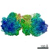
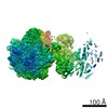
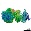
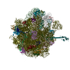
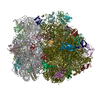
single particle reconstruction / cryo EM / Resolution: 4.92 Å
Journal: Nature / Year: 2022 Title: Bacterial ribosome collision sensing by a MutS DNA repair ATPase paralogue. Authors: Federico Cerullo / Sebastian Filbeck / Pratik Rajendra Patil / Hao-Chih Hung / Haifei Xu / Julia Vornberger / Florian W Hofer / Jaro Schmitt / Guenter Kramer / Bernd Bukau / Kay Hofmann / ...Authors: Federico Cerullo / Sebastian Filbeck / Pratik Rajendra Patil / Hao-Chih Hung / Haifei Xu / Julia Vornberger / Florian W Hofer / Jaro Schmitt / Guenter Kramer / Bernd Bukau / Kay Hofmann / Stefan Pfeffer / Claudio A P Joazeiro / Abstract: Ribosome stalling during translation is detrimental to cellular fitness, but how this is sensed and elicits recycling of ribosomal subunits and quality control of associated mRNA and incomplete ...Ribosome stalling during translation is detrimental to cellular fitness, but how this is sensed and elicits recycling of ribosomal subunits and quality control of associated mRNA and incomplete nascent chains is poorly understood. Here we uncover Bacillus subtilis MutS2, a member of the conserved MutS family of ATPases that function in DNA mismatch repair, as an unexpected ribosome-binding protein with an essential function in translational quality control. Cryo-electron microscopy analysis of affinity-purified native complexes shows that MutS2 functions in sensing collisions between stalled and translating ribosomes and suggests how ribosome collisions can serve as platforms to deploy downstream processes: MutS2 has an RNA endonuclease small MutS-related (SMR) domain, as well as an ATPase/clamp domain that is properly positioned to promote ribosomal subunit dissociation, which is a requirement both for ribosome recycling and for initiation of ribosome-associated protein quality control (RQC). Accordingly, MutS2 promotes nascent chain modification with alanine-tail degrons-an early step in RQC-in an ATPase domain-dependent manner. The relevance of these observations is underscored by evidence of strong co-occurrence of MutS2 and RQC genes across bacterial phyla. Overall, the findings demonstrate a deeply conserved role for ribosome collisions in mounting a complex response to the interruption of translation within open reading frames.
History
Deposition
Jan 19, 2022
-
Header (metadata) release
Mar 9, 2022
-
Map release
Mar 9, 2022
-
Update
Dec 13, 2023
-
Current status
Dec 13, 2023
Processing site: PDBe / Status: Released
-
Structure visualization
Movie
Surface view with section colored by density value
In the structure databanks used in Yorodumi, some data are registered as the other names, "COVID-19 virus" and "2019-nCoV". Here are the details of the virus and the list of structure data.
Jan 31, 2019. EMDB accession codes are about to change! (news from PDBe EMDB page)
EMDB accession codes are about to change! (news from PDBe EMDB page)
The allocation of 4 digits for EMDB accession codes will soon come to an end. Whilst these codes will remain in use, new EMDB accession codes will include an additional digit and will expand incrementally as the available range of codes is exhausted. The current 4-digit format prefixed with “EMD-” (i.e. EMD-XXXX) will advance to a 5-digit format (i.e. EMD-XXXXX), and so on. It is currently estimated that the 4-digit codes will be depleted around Spring 2019, at which point the 5-digit format will come into force.
The EM Navigator/Yorodumi systems omit the EMD- prefix.
Related info.:Q: What is EMD? / ID/Accession-code notation in Yorodumi/EM Navigator
Yorodumi is a browser for structure data from EMDB, PDB, SASBDB, etc.
This page is also the successor to EM Navigator detail page, and also detail information page/front-end page for Omokage search.
The word "yorodu" (or yorozu) is an old Japanese word meaning "ten thousand". "mi" (miru) is to see.
Related info.:EMDB / PDB / SASBDB / Comparison of 3 databanks / Yorodumi Search / Aug 31, 2016. New EM Navigator & Yorodumi / Yorodumi Papers / Jmol/JSmol / Function and homology information / Changes in new EM Navigator and Yorodumi
 Movie
Movie Controller

 Yorodumi
 Open data
 Basic information
 Map data
 Sample
 Keywords
 Collision / MutS2 / disome / RQC /
  Function and homology information
 Authors
 Germany, 1 items
  Citation
 Structure visualization
 Downloads & links
 emd_14161.png
 http://ftp.pdbj.org/pub/emdb/structures/EMD-14161


 Sample components
 Processing
 Electron microscopy
