Biotechnology and Biological Sciences Research Council (BBSRC)
BB/S003339/1
United Kingdom
Medical Research Council (MRC, United Kingdom)
MR/V000403/1
United Kingdom
Citation
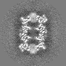
Journal: Structure / Year: 2022 Title: IceBreaker: Software for high-resolution single-particle cryo-EM with non-uniform ice. Authors: Mateusz Olek / Kevin Cowtan / Donovan Webb / Yuriy Chaban / Peijun Zhang / Abstract: Despite the abundance of available software tools, optimal particle selection is still a vital issue in single-particle cryoelectron microscopy (cryo-EM). Regardless of the method used, most pickers ...Despite the abundance of available software tools, optimal particle selection is still a vital issue in single-particle cryoelectron microscopy (cryo-EM). Regardless of the method used, most pickers struggle when ice thickness varies on a micrograph. IceBreaker allows users to estimate the relative ice gradient and flatten it by equalizing the local contrast. It allows the differentiation of particles from the background and improves overall particle picking performance. Furthermore, we introduce an additional parameter corresponding to local ice thickness for each particle. Particles with a defined ice thickness can be grouped and filtered based on this parameter during processing. These functionalities are especially valuable for on-the-fly processing to automatically pick as many particles as possible from each micrograph and to select optimal regions for data collection. Finally, estimated ice gradient distributions can be stored separately and used to inspect the quality of prepared samples.
In the structure databanks used in Yorodumi, some data are registered as the other names, "COVID-19 virus" and "2019-nCoV". Here are the details of the virus and the list of structure data.
Jan 31, 2019. EMDB accession codes are about to change! (news from PDBe EMDB page)
EMDB accession codes are about to change! (news from PDBe EMDB page)
The allocation of 4 digits for EMDB accession codes will soon come to an end. Whilst these codes will remain in use, new EMDB accession codes will include an additional digit and will expand incrementally as the available range of codes is exhausted. The current 4-digit format prefixed with “EMD-” (i.e. EMD-XXXX) will advance to a 5-digit format (i.e. EMD-XXXXX), and so on. It is currently estimated that the 4-digit codes will be depleted around Spring 2019, at which point the 5-digit format will come into force.
The EM Navigator/Yorodumi systems omit the EMD- prefix.
Related info.:Q: What is EMD? / ID/Accession-code notation in Yorodumi/EM Navigator
Yorodumi is a browser for structure data from EMDB, PDB, SASBDB, etc.
This page is also the successor to EM Navigator detail page, and also detail information page/front-end page for Omokage search.
The word "yorodu" (or yorozu) is an old Japanese word meaning "ten thousand". "mi" (miru) is to see.
Related info.:EMDB / PDB / SASBDB / Comparison of 3 databanks / Yorodumi Search / Aug 31, 2016. New EM Navigator & Yorodumi / Yorodumi Papers / Jmol/JSmol / Function and homology information / Changes in new EM Navigator and Yorodumi
 Movie
Movie Controller

 Yorodumi
 Open data
 Basic information
 Map data
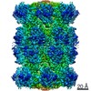
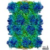
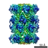
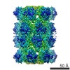
 Sample
 Function and homology information
 proteasome endopeptidase complex / proteasome core complex, beta-subunit complex / proteasome core complex, alpha-subunit complex / threonine-type endopeptidase activity / proteasomal protein catabolic process / ubiquitin-dependent protein catabolic process /

 Authors
 United Kingdom, 3 items
  Citation
 Structure visualization
 Downloads & links
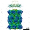
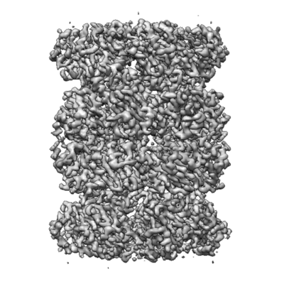 emd_13312.png
 http://ftp.pdbj.org/pub/emdb/structures/EMD-13312


 Z
Z Y
Y X
X


 Sample components
 Processing
 Electron microscopy

