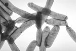
Journal: mBio / Year: 2021 Title: The Polar Icm/Dot T4SS Establishes Distinct Contact Sites with the Pathogen Vacuole Membrane. Authors: Désirée Böck / Dario Hüsler / Bernhard Steiner / João M Medeiros / Amanda Welin / Katarzyna A Radomska / Wolf-Dietrich Hardt / Martin Pilhofer / Hubert Hilbi / Abstract: Legionella pneumophila, the causative agent of Legionnaires' disease, is a facultative intracellular pathogen that survives inside phagocytic host cells by establishing a protected replication niche, ...Legionella pneumophila, the causative agent of Legionnaires' disease, is a facultative intracellular pathogen that survives inside phagocytic host cells by establishing a protected replication niche, termed the "-containing vacuole" (LCV). To form an LCV and subvert pivotal host pathways, L. pneumophila employs a type IV secretion system (T4SS), which translocates more than 300 different effector proteins into the host cell. The L. pneumophila T4SS complex has been shown to span the bacterial cell envelope at the bacterial poles. However, the interactions between the T4SS and the LCV membrane are not understood. Using cryo-focused ion beam milling, cryo-electron tomography, and confocal laser scanning fluorescence microscopy, we show that up to half of the intravacuolar L. pneumophila bacteria tether their cell pole to the LCV membrane. Tethering coincides with the presence and function of T4SSs and likely promotes the establishment of distinct contact sites between T4SSs and the LCV membrane. Contact sites are characterized by indentations in the limiting LCV membrane and localize juxtaposed to T4SS machineries. The data are in agreement with the notion that effector translocation occurs by close membrane contact rather than by an extended pilus. Our findings provide novel insights into the interactions of the L. pneumophila T4SS with the LCV membrane . Legionnaires' disease is a life-threatening pneumonia, which is characterized by high fever, coughing, shortness of breath, muscle pain, and headache. The disease is caused by the amoeba-resistant bacterium L. pneumophila found in various soil and aquatic environments and is transmitted to humans via the inhalation of small bacteria-containing droplets. An essential virulence factor of L. pneumophila is a so-called "type IV secretion system" (T4SS), which, by injecting a plethora of "effector proteins" into the host cell, determines pathogen-host interactions and the formation of a distinct intracellular compartment, the "-containing vacuole" (LCV). It is unknown how the T4SS makes contact to the LCV membrane to deliver the effectors. In this study, we identify indentations in the host cell membrane in close proximity to functional T4SSs localizing at the bacterial poles. Our work reveals first insights into the architecture of -LCV contact sites.
Cryogen name: ETHANE-PROPANE / Chamber humidity: 90 % / Chamber temperature: 280 K / Instrument: FEI VITROBOT MARK IV
Details
cryoFIB milling of plunge-frozen infected amoeba and subsequent cryoET of the resulting lamellae
Sectioning
Focused ion beam - Instrument: OTHER / Focused ion beam - Ion: OTHER / Focused ion beam - Voltage: 30 kV / Focused ion beam - Current: 0.025 nA / Focused ion beam - Duration: 3600 sec. / Focused ion beam - Temperature: 120 K / Focused ion beam - Initial thickness: 1000 nm / Focused ion beam - Final thickness: 200 nm Focused ion beam - Details: The value given for _emd_sectioning_focused_ion_beam.instrument is FEI Helios NanoLab600i. This is not in a list of allowed values {'OTHER', 'DB235'} so OTHER is written into the XML file.
-
Electron microscopy
Microscope
FEI TITAN KRIOS
Electron beam
Acceleration voltage: 300 kV / Electron source: FIELD EMISSION GUN
In the structure databanks used in Yorodumi, some data are registered as the other names, "COVID-19 virus" and "2019-nCoV". Here are the details of the virus and the list of structure data.
Jan 31, 2019. EMDB accession codes are about to change! (news from PDBe EMDB page)
EMDB accession codes are about to change! (news from PDBe EMDB page)
The allocation of 4 digits for EMDB accession codes will soon come to an end. Whilst these codes will remain in use, new EMDB accession codes will include an additional digit and will expand incrementally as the available range of codes is exhausted. The current 4-digit format prefixed with “EMD-” (i.e. EMD-XXXX) will advance to a 5-digit format (i.e. EMD-XXXXX), and so on. It is currently estimated that the 4-digit codes will be depleted around Spring 2019, at which point the 5-digit format will come into force.
The EM Navigator/Yorodumi systems omit the EMD- prefix.
Related info.:Q: What is EMD? / ID/Accession-code notation in Yorodumi/EM Navigator
Yorodumi is a browser for structure data from EMDB, PDB, SASBDB, etc.
This page is also the successor to EM Navigator detail page, and also detail information page/front-end page for Omokage search.
The word "yorodu" (or yorozu) is an old Japanese word meaning "ten thousand". "mi" (miru) is to see.
Related info.:EMDB / PDB / SASBDB / Comparison of 3 databanks / Yorodumi Search / Aug 31, 2016. New EM Navigator & Yorodumi / Yorodumi Papers / Jmol/JSmol / Function and homology information / Changes in new EM Navigator and Yorodumi
 Movie
Movie Controller

 Yorodumi
 Open data
 Basic information
 Map data
 Sample
 Legionella pneumophila (bacteria)
 Authors
 Switzerland, 2 items
  Citation
 Structure visualization
 Movie viewer
 Downloads & links
 emd_13246.png
 http://ftp.pdbj.org/pub/emdb/structures/EMD-13246

 Sample components
 Processing
 Electron microscopy
