 Movie
Movie Controller


+ Open data

- Basic information
| Entry | Database: EMDB / ID: EMD-13153 | |||||||||||||||
|---|---|---|---|---|---|---|---|---|---|---|---|---|---|---|---|---|
| Title | 2.43 A Mycobacterium marinum EspB. | |||||||||||||||
 Map data Map data | Local B-factor sharpened map from LocSpiral | |||||||||||||||
 Sample Sample |
| |||||||||||||||
| Function / homology | ESX-1 secretion-associated protein EspB, PE domain / ESX-1 secreted protein B PE domain / extracellular region / ESX-1 secretion-associated protein EspB Function and homology information Function and homology information | |||||||||||||||
| Biological species |   Mycobacterium marinum (bacteria) / Mycobacterium marinum (strain ATCC BAA-535 / M) (bacteria) Mycobacterium marinum (bacteria) / Mycobacterium marinum (strain ATCC BAA-535 / M) (bacteria) | |||||||||||||||
| Method | single particle reconstruction / cryo EM / Resolution: 2.43 Å | |||||||||||||||
 Authors Authors | Gijsbers A / Zhang Y / Vinciauskaite V / Siroy A / Ye G / Tria G / Mathew A / Sanchez-Puig N / Lopez-Iglesias C / Peters PJ / Ravelli RBG | |||||||||||||||
| Funding support |  Netherlands, European Union, Netherlands, European Union,  Mexico, 4 items Mexico, 4 items
| |||||||||||||||
 Citation Citation | Journal: Curr Res Struct Biol / Year: 2021 Title: Priming mycobacterial ESX-secreted protein B to form a channel-like structure. Authors: Abril Gijsbers / Vanesa Vinciauskaite / Axel Siroy / Ye Gao / Giancarlo Tria / Anjusha Mathew / Nuria Sánchez-Puig / Carmen López-Iglesias / Peter J Peters / Raimond B G Ravelli / Abstract: ESX-1 is a major virulence factor of , a secretion machinery directly involved in the survival of the microorganism from the immune system defence. It disrupts the phagosome membrane of the host cell ...ESX-1 is a major virulence factor of , a secretion machinery directly involved in the survival of the microorganism from the immune system defence. It disrupts the phagosome membrane of the host cell through a contact-dependent mechanism. Recently, the structure of the inner-membrane core complex of the homologous ESX-3 and ESX-5 was resolved; however, the elements involved in the secretion through the outer membrane or those acting on the host cell membrane are unknown. Protein substrates might form this missing element. Here, we describe the oligomerisation process of the ESX-1 substrate EspB, which occurs upon cleavage of its C-terminal region and is favoured by an acidic environment. Cryo-electron microscopy data shows that quaternary structure of EspB is conserved across slow growing species, but not in the fast growing . EspB assembles into a channel with dimensions and characteristics suitable for the transit of ESX-1 substrates, as shown by the presence of another EspB trapped within. Our results provide insight into the structure and assembly of EspB, and suggests a possible function as a structural element of ESX-1. | |||||||||||||||
| History |
|
- Structure visualization
| Movie |
Movie viewer |
|---|---|
| Structure viewer | EM map: SurfViewMolmilJmol/JSmol |
| Supplemental images |

- Downloads & links
-EMDB archive
| Map data | emd_13153.map.gz | 9.3 MB | EMDB map data format | |
|---|---|---|---|---|
| Header (meta data) | emd-13153-v30.xmlemd-13153.xml | 22.6 KB 22.6 KB | Display Display | EMDB header |
| FSC (resolution estimation) | emd_13153_fsc.xml | 9.1 KB | Display | FSC data file |
| Images | 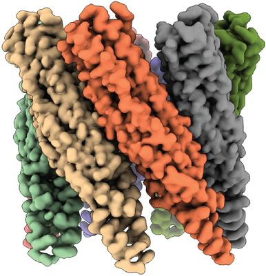 emd_13153.png emd_13153.png | 210.4 KB | ||
| Masks | emd_13153_msk_1.map | 64 MB | Mask map | |
| Others | emd_13153_additional_1.map.gzemd_13153_half_map_1.map.gzemd_13153_half_map_2.map.gz | 6.8 MB 49.5 MB 49.6 MB | ||
| Archive directory |  http://ftp.pdbj.org/pub/emdb/structures/EMD-13153ftp://ftp.pdbj.org/pub/emdb/structures/EMD-13153 http://ftp.pdbj.org/pub/emdb/structures/EMD-13153ftp://ftp.pdbj.org/pub/emdb/structures/EMD-13153 | HTTPS FTP |
-Related structure data
| Related structure data |  7p0zMC  7p13C M: atomic model generated by this map C: citing same article ( |
|---|---|
| Similar structure data |


-Links
| EMDB pages | EMDB (EBI/PDBe) / EMDataResource |
|---|
-Map
| File | Download / File: emd_13153.map.gz / Format: CCP4 / Size: 64 MB / Type: IMAGE STORED AS FLOATING POINT NUMBER (4 BYTES) | ||||||||||||||||||||||||||||||||||||||||||||||||||||||||||||||||||||
|---|---|---|---|---|---|---|---|---|---|---|---|---|---|---|---|---|---|---|---|---|---|---|---|---|---|---|---|---|---|---|---|---|---|---|---|---|---|---|---|---|---|---|---|---|---|---|---|---|---|---|---|---|---|---|---|---|---|---|---|---|---|---|---|---|---|---|---|---|---|
| Annotation | Local B-factor sharpened map from LocSpiral | ||||||||||||||||||||||||||||||||||||||||||||||||||||||||||||||||||||
| Voxel size | X=Y=Z: 0.834 Å | ||||||||||||||||||||||||||||||||||||||||||||||||||||||||||||||||||||
| Density |
| ||||||||||||||||||||||||||||||||||||||||||||||||||||||||||||||||||||
| Symmetry | Space group: 1 | ||||||||||||||||||||||||||||||||||||||||||||||||||||||||||||||||||||
| Details | EMDB XML:
CCP4 map header:
| ||||||||||||||||||||||||||||||||||||||||||||||||||||||||||||||||||||
-Supplemental data
-Mask #1
| File | emd_13153_msk_1.map | ||||||||||||
|---|---|---|---|---|---|---|---|---|---|---|---|---|---|
| Projections & Slices |
| ||||||||||||
| Density Histograms |
 Z
Z Y
Y X
X


-Additional map: Global b-factor sharpened map from Relion postprocess.
| File | emd_13153_additional_1.map | ||||||||||||
|---|---|---|---|---|---|---|---|---|---|---|---|---|---|
| Annotation | Global b-factor sharpened map from Relion postprocess. | ||||||||||||
| Projections & Slices |
| ||||||||||||
| Density Histograms |


-Half map: Unmasked halfmap 1 from Refine3D
| File | emd_13153_half_map_1.map | ||||||||||||
|---|---|---|---|---|---|---|---|---|---|---|---|---|---|
| Annotation | Unmasked halfmap 1 from Refine3D | ||||||||||||
| Projections & Slices |
| ||||||||||||
| Density Histograms |


-Half map: Unmasked halfmap 2 from Refine3D
| File | emd_13153_half_map_2.map | ||||||||||||
|---|---|---|---|---|---|---|---|---|---|---|---|---|---|
| Annotation | Unmasked halfmap 2 from Refine3D | ||||||||||||
| Projections & Slices |
| ||||||||||||
| Density Histograms |


- Sample components
-Entire : Heptamer of EspB
| Entire | Name: Heptamer of EspB |
|---|---|
| Components |
|
-Supramolecule #1: Heptamer of EspB
| Supramolecule | Name: Heptamer of EspB / type: complex / ID: 1 / Parent: 0 / Macromolecule list: all |
|---|---|
| Source (natural) | Organism: Mycobacterium marinum (bacteria) / Strain: BAA-535 |
| Recombinant expression | Organism: Escherichia coli (E. coli) / Recombinant plasmid: pAG10 |
| Molecular weight | Experimental: 210 KDa |
-Macromolecule #1: ESX-1 secretion-associated protein EspB
| Macromolecule | Name: ESX-1 secretion-associated protein EspB / type: protein_or_peptide / ID: 1 / Number of copies: 7 / Enantiomer: LEVO |
|---|---|
| Source (natural) | Organism: Mycobacterium marinum (strain ATCC BAA-535 / M) (bacteria) Strain: ATCC BAA-535 / M |
| Molecular weight | Theoretical: 31.274275 KDa |
| Recombinant expression | Organism: Escherichia coli (E. coli) |
| Sequence | String: SMHSQPQTVT VDQQEILNRA DEVEAPMATP PTDVPQAPSG LTAANNAAEQ LAVSADNVRL YLQAGERERQ RLATSLRNAA AAYGEVEDE SATALDNDGN GEVDAQSAGG AGAGQTESLE ETPKVAAAGE SDFTDLKTAA TKLESGDQGT SMVNFADGWN N FNLSLQRD ...String: SMHSQPQTVT VDQQEILNRA DEVEAPMATP PTDVPQAPSG LTAANNAAEQ LAVSADNVRL YLQAGERERQ RLATSLRNAA AAYGEVEDE SATALDNDGN GEVDAQSAGG AGAGQTESLE ETPKVAAAGE SDFTDLKTAA TKLESGDQGT SMVNFADGWN N FNLSLQRD IKRFRIFENW EGDAATACEA SMDQQKEWIL HMAKLSASLA KQANFMAQLQ LWARRGHPTL ADIVELERLA KD PDYQEQA IKLYAEYQET SEKVLSEYNT KADLEPVNPP KPPAAIKIDP P |
-Experimental details
-Structure determination
| Method | cryo EM |
|---|---|
 Processing Processing | single particle reconstruction |
| Aggregation state | particle |
-Sample preparation
| Concentration | 8.7 mg/mL | |||||||||
|---|---|---|---|---|---|---|---|---|---|---|
| Buffer | pH: 5.5 Component:
| |||||||||
| Grid | Model: UltrAuFoil R1.2/1.3 / Material: GOLD / Mesh: 300 / Support film - Material: GOLD / Support film - topology: HOLEY ARRAY / Support film - Film thickness: 50.0 nm / Pretreatment - Type: GLOW DISCHARGE / Pretreatment - Atmosphere: AIR | |||||||||
| Vitrification | Cryogen name: ETHANE / Chamber humidity: 100 % / Chamber temperature: 277 K / Instrument: FEI VITROBOT MARK IV | |||||||||
| Details | This sample was monodisperse. |
- Electron microscopy
| Microscope | TFS KRIOS |
|---|---|
| Electron beam | Acceleration voltage: 300 kV / Electron source: FIELD EMISSION GUN |
| Electron optics | C2 aperture diameter: 50.0 µm / Calibrated magnification: 105000 / Illumination mode: FLOOD BEAM / Imaging mode: BRIGHT FIELDBright-field microscopy / Cs: 2.7 mm / Nominal defocus max: -2.0 µm / Nominal defocus min: -1.25 µm / Nominal magnification: 105000 |
| Specialist optics | Energy filter - Name: GIF Bioquantum / Energy filter - Slit width: 20 eV |
| Sample stage | Specimen holder model: FEI TITAN KRIOS AUTOGRID HOLDER / Cooling holder cryogen: NITROGEN |
| Temperature | Min: 90.0 K |
| Details | Basic direct alignments were done as well as astigmatism and coma alignment using AutoCTF |
| Image recording | Film or detector model: GATAN K3 BIOQUANTUM (6k x 4k) / Digitization - Dimensions - Width: 5760 pixel / Digitization - Dimensions - Height: 4092 pixel / Digitization - Sampling interval: 5.0 µm / Number grids imaged: 1 / Number real images: 2421 / Average exposure time: 1.8 sec. / Average electron dose: 40.0 e/Å2 |
| Experimental equipment |  Model: Titan Krios / Image courtesy: FEI Company |
-Image processing
| CTF correction | Software - Name: Gctf (ver. 1.06) |
|---|---|
| Startup model | Type of model: PDB ENTRY PDB model - PDB ID: |
| Initial angle assignment | Type: MAXIMUM LIKELIHOOD / Software - Name: RELION (ver. 3.1) |
| Final angle assignment | Type: MAXIMUM LIKELIHOOD / Software - Name: RELION (ver. 3.1) |
| Final reconstruction | Applied symmetry - Point group: C7 (7 fold cyclic) / Algorithm: FOURIER SPACE / Resolution.type: BY AUTHOR / Resolution: 2.43 Å / Resolution method: FSC 0.143 CUT-OFF / Software - Name: RELION (ver. 3.1) / Number images used: 435505 |
| FSC plot (resolution estimation) |  |

