 Movie
Movie Controller


+ Open data

- Basic information
| Entry | Database: EMDB / ID: EMD-13087 | |||||||||||||||||||||||||||
|---|---|---|---|---|---|---|---|---|---|---|---|---|---|---|---|---|---|---|---|---|---|---|---|---|---|---|---|---|
| Title | Immature HIV-1 matrix structure | |||||||||||||||||||||||||||
 Map data Map data | Immature HIV-1 MA structure | |||||||||||||||||||||||||||
 Sample Sample |
| |||||||||||||||||||||||||||
| Function / homology |  Function and homology information Function and homology informationviral process / viral nucleocapsid / host cell cytoplasm / virion membrane / structural molecule activity /  RNA binding / zinc ion binding / cytoplasm RNA binding / zinc ion binding / cytoplasmSimilarity search - Function | |||||||||||||||||||||||||||
| Biological species |  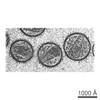 Human immunodeficiency virus 1 Human immunodeficiency virus 1 | |||||||||||||||||||||||||||
| Method | subtomogram averaging / cryo EM / Resolution: 7.2 Å | |||||||||||||||||||||||||||
 Authors Authors | Qu K / Ke ZL / Zila V / Anders-Oesswein M / Glass B / Muecksch F / Mueller R / Schultz C / Mueller B / Kraeusslich HG / Briggs JAG | |||||||||||||||||||||||||||
| Funding support |  Germany, 8 items Germany, 8 items
| |||||||||||||||||||||||||||
 Citation Citation | Journal: Science / Year: 2021 Title: Maturation of the matrix and viral membrane of HIV-1. Authors: Kun Qu / Zunlong Ke / Vojtech Zila / Maria Anders-Össwein / Bärbel Glass / Frauke Mücksch / Rainer Müller / Carsten Schultz / Barbara Müller / Hans-Georg Kräusslich / John A G Briggs /   Abstract: Gag, the primary structural protein of HIV-1, is recruited to the plasma membrane for virus assembly by its matrix (MA) domain. Gag is subsequently cleaved into its component domains, causing ...Gag, the primary structural protein of HIV-1, is recruited to the plasma membrane for virus assembly by its matrix (MA) domain. Gag is subsequently cleaved into its component domains, causing structural maturation to repurpose the virion for cell entry. We determined the structure and arrangement of MA within immature and mature HIV-1 through cryo-electron tomography. We found that MA rearranges between two different hexameric lattices upon maturation. In mature HIV-1, a lipid extends out of the membrane to bind with a pocket in MA. Our data suggest that proteolytic maturation of HIV-1 not only assembles the viral capsid surrounding the genome but also repurposes the membrane-bound MA lattice for an entry or postentry function and results in the partial removal of up to 2500 lipids from the viral membrane. | |||||||||||||||||||||||||||
| History |
|
- Structure visualization
| Movie |
Movie viewer |
|---|---|
| Structure viewer | EM map: SurfViewMolmilJmol/JSmol |
| Supplemental images |

- Downloads & links
-EMDB archive
| Map data | emd_13087.map.gz | 17.2 MB | EMDB map data format | |
|---|---|---|---|---|
| Header (meta data) | emd-13087-v30.xmlemd-13087.xml | 14.5 KB 14.5 KB | Display Display | EMDB header |
| Images | 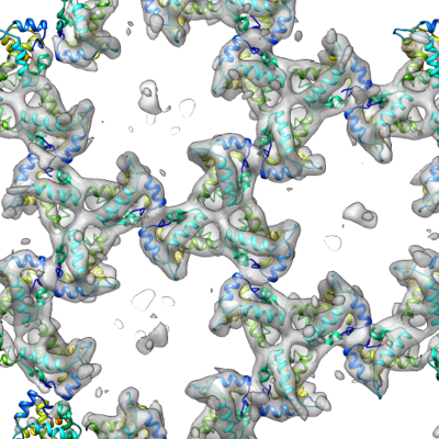 emd_13087.png emd_13087.png | 217.6 KB | ||
| Archive directory |  http://ftp.pdbj.org/pub/emdb/structures/EMD-13087ftp://ftp.pdbj.org/pub/emdb/structures/EMD-13087 http://ftp.pdbj.org/pub/emdb/structures/EMD-13087ftp://ftp.pdbj.org/pub/emdb/structures/EMD-13087 | HTTPS FTP |
-Related structure data
| Related structure data |  7ovqMC 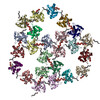 7ovrC M: atomic model generated by this map C: citing same article ( |
|---|---|
| Similar structure data |


-Links
| EMDB pages | EMDB (EBI/PDBe) / EMDataResource |
|---|---|
| Related items in Molecule of the Month |

-Map
| File | Download / File: emd_13087.map.gz / Format: CCP4 / Size: 27 MB / Type: IMAGE STORED AS FLOATING POINT NUMBER (4 BYTES) | ||||||||||||||||||||||||||||||||||||||||||||||||||||||||||||||||||||
|---|---|---|---|---|---|---|---|---|---|---|---|---|---|---|---|---|---|---|---|---|---|---|---|---|---|---|---|---|---|---|---|---|---|---|---|---|---|---|---|---|---|---|---|---|---|---|---|---|---|---|---|---|---|---|---|---|---|---|---|---|---|---|---|---|---|---|---|---|---|
| Annotation | Immature HIV-1 MA structure | ||||||||||||||||||||||||||||||||||||||||||||||||||||||||||||||||||||
| Voxel size | X=Y=Z: 1.35 Å | ||||||||||||||||||||||||||||||||||||||||||||||||||||||||||||||||||||
| Density |
| ||||||||||||||||||||||||||||||||||||||||||||||||||||||||||||||||||||
| Symmetry | Space group: 1 | ||||||||||||||||||||||||||||||||||||||||||||||||||||||||||||||||||||
| Details | EMDB XML:
CCP4 map header:
| ||||||||||||||||||||||||||||||||||||||||||||||||||||||||||||||||||||
-Supplemental data
- Sample components
-Entire : Human immunodeficiency virus 1
| Entire | Name: Human immunodeficiency virus 1 |
|---|---|
| Components |
|
-Supramolecule #1: Human immunodeficiency virus 1
| Supramolecule | Name: Human immunodeficiency virus 1 / type: virus / ID: 1 / Parent: 0 / Macromolecule list: #1 Details: HIV-1 NL4-3 protease defective (D25A) virus particles purified from HEK293T cells. NCBI-ID: 11676 / Sci species name: Human immunodeficiency virus 1 / Virus type: VIRION / Virus isolate: STRAIN / Virus enveloped: Yes / Virus empty: No |
|---|---|
| Host system | Organism:  Homo sapiens (human) Homo sapiens (human) |
-Macromolecule #1: Gag polyprotein
| Macromolecule | Name: Gag polyprotein / type: protein_or_peptide / ID: 1 / Number of copies: 24 / Enantiomer: LEVO |
|---|---|
| Source (natural) | Organism: Human immunodeficiency virus 1 |
| Molecular weight | Theoretical: 13.084983 KDa |
| Recombinant expression | Organism: Homo sapiens (human) |
| Sequence | String: GARASVLSGG ELDKWEKIRL RPGGKKQYKL KHIVWASREL ERFAVNPGLL ETSEGCRQIL GQLQPSLQTG SEELRSLYNT IAVLYCVHQ RIDVKDTKEA LDKIEEEQNK SKKKAQ |
-Macromolecule #2: MYRISTIC ACID
| Macromolecule | Name: MYRISTIC ACID / type: ligand / ID: 2 / Number of copies: 24 / Formula: MYR |
|---|---|
| Molecular weight | Theoretical: 228.371 Da |
| Chemical component information |  ChemComp-MYR: |
-Experimental details
-Structure determination
| Method | cryo EM |
|---|---|
 Processing Processing | subtomogram averaging |
| Aggregation state | particle |
-Sample preparation
| Buffer | pH: 7.4 |
|---|---|
| Vitrification | Cryogen name: ETHANE |
| Details | HIV-1 NL4-3 protease defective (D25A) virus particles purified from HEK293T cells. |
- Electron microscopy
| Microscope | FEI TITAN KRIOS |
|---|---|
| Electron beam | Acceleration voltage: 300 kV / Electron source: FIELD EMISSION GUN |
| Electron optics | C2 aperture diameter: 50.0 µm / Illumination mode: FLOOD BEAM / Imaging mode: BRIGHT FIELDBright-field microscopy / Cs: 2.7 mm / Nominal defocus max: 5.0 µm / Nominal defocus min: 1.5 µm / Nominal magnification: 105000 |
| Specialist optics | Energy filter - Name: GIF Quantum LS / Energy filter - Slit width: 20 eV |
| Sample stage | Specimen holder model: FEI TITAN KRIOS AUTOGRID HOLDER / Cooling holder cryogen: NITROGEN |
| Image recording | Film or detector model: GATAN K2 QUANTUM (4k x 4k) / Detector mode: SUPER-RESOLUTION / Average electron dose: 3.0 e/Å2 / Details: Number of frames ranged from 10-12. |
| Experimental equipment |  Model: Titan Krios / Image courtesy: FEI Company |
-Image processing
| Extraction | Number tomograms: 74 / Number images used: 251207 / Software - Name: MATLAB |
|---|---|
| CTF correction | Software: (Name: CTFFIND, NOVACTF) |
| Final angle assignment | Type: ANGULAR RECONSTITUTION |
| Final reconstruction | Applied symmetry - Point group: C3 (3 fold cyclic) / Resolution.type: BY AUTHOR / Resolution: 7.2 Å / Resolution method: FSC 0.143 CUT-OFF / Software - Name: AV3 / Number subtomograms used: 22417 |

