Netherlands Organisation for Scientific Research (NWO)
NWO.STU.018-2.007
Netherlands
Citation
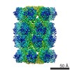
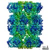
Journal: Elife / Year: 2022 Title: Nanofluidic chips for cryo-EM structure determination from picoliter sample volumes. Authors: Stefan T Huber / Edin Sarajlic / Roeland Huijink / Felix Weis / Wiel H Evers / Arjen J Jakobi / Abstract: Cryogenic electron microscopy has become an essential tool for structure determination of biological macromolecules. In practice, the difficulty to reliably prepare samples with uniform ice thickness ...Cryogenic electron microscopy has become an essential tool for structure determination of biological macromolecules. In practice, the difficulty to reliably prepare samples with uniform ice thickness still represents a barrier for routine high-resolution imaging and limits the current throughput of the technique. We show that a nanofluidic sample support with well-defined geometry can be used to prepare cryo-EM specimens with reproducible ice thickness from picoliter sample volumes. The sample solution is contained in electron-transparent nanochannels that provide uniform thickness gradients without further optimisation and eliminate the potentially destructive air-water interface. We demonstrate the possibility to perform high-resolution structure determination with three standard protein specimens. Nanofabricated sample supports bear potential to automate the cryo-EM workflow, and to explore new frontiers for cryo-EM applications such as time-resolved imaging and high-throughput screening.
EMPIAR-10708 (Title: Apoferritin, TMV and T20S proteasome in nanofluidic channels Data size: 308.3 Data #1: Unaligned multi-frame movies of pyrococcus furiosus apoferritin in silicon nitride nanochannels [micrographs - multiframe] Data #2: Multi-frame movies of T20S proteasome in silicon nitride nanochannels [micrographs - multiframe] Data #3: Multi-frame movies of TMV in silicon nitride nanochannels [micrographs - multiframe])
Cryogen name: ETHANE / Instrument: LEICA PLUNGER Details: The sample was filled into cryoChips through the cantilever and then transferred within ~10 seconds to the Leica plunger for freezing..
Details
The sample was filled into nanofluidic channels.
-
Electron microscopy
Microscope
JEOL 3200FSC
Electron beam
Acceleration voltage: 300 kV / Electron source: FIELD EMISSION GUN
Film or detector model: GATAN K2 SUMMIT (4k x 4k) / Detector mode: COUNTING / Number grids imaged: 1 / Number real images: 121 / Average exposure time: 11.0 sec. / Average electron dose: 59.7 e/Å2
-
Image processing
CTF correction
Software - Name: cryoSPARC (ver. 3.1)
Startup model
Type of model: OTHER / Details: Stochastic gradient decent in cryoSPARC 3.1
Initial angle assignment
Type: NOT APPLICABLE
Final 3D classification
Number classes: 1 / Software - Name: cryoSPARC (ver. 3.1)
Final angle assignment
Type: MAXIMUM LIKELIHOOD / Software - Name: cryoSPARC (ver. 3.1)
Final reconstruction
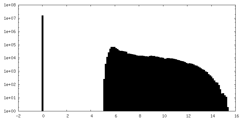
Number classes used: 1 / Applied symmetry - Point group: D7 (2x7 fold dihedral) / Algorithm: FOURIER SPACE / Resolution.type: BY AUTHOR / Resolution: 5.4 Å / Resolution method: FSC 0.143 CUT-OFF / Software - Name: cryoSPARC (ver. 3.1) / Number images used: 5750
FSC plot (resolution estimation)
+
About Yorodumi
-
News
-
Feb 9, 2022. New format data for meta-information of EMDB entries
New format data for meta-information of EMDB entries
Version 3 of the EMDB header file is now the official format.
The previous official version 1.9 will be removed from the archive.
In the structure databanks used in Yorodumi, some data are registered as the other names, "COVID-19 virus" and "2019-nCoV". Here are the details of the virus and the list of structure data.
Jan 31, 2019. EMDB accession codes are about to change! (news from PDBe EMDB page)
EMDB accession codes are about to change! (news from PDBe EMDB page)
The allocation of 4 digits for EMDB accession codes will soon come to an end. Whilst these codes will remain in use, new EMDB accession codes will include an additional digit and will expand incrementally as the available range of codes is exhausted. The current 4-digit format prefixed with “EMD-” (i.e. EMD-XXXX) will advance to a 5-digit format (i.e. EMD-XXXXX), and so on. It is currently estimated that the 4-digit codes will be depleted around Spring 2019, at which point the 5-digit format will come into force.
The EM Navigator/Yorodumi systems omit the EMD- prefix.
Related info.:Q: What is EMD? / ID/Accession-code notation in Yorodumi/EM Navigator
Yorodumi is a browser for structure data from EMDB, PDB, SASBDB, etc.
This page is also the successor to EM Navigator detail page, and also detail information page/front-end page for Omokage search.
The word "yorodu" (or yorozu) is an old Japanese word meaning "ten thousand". "mi" (miru) is to see.
Related info.:EMDB / PDB / SASBDB / Comparison of 3 databanks / Yorodumi Search / Aug 31, 2016. New EM Navigator & Yorodumi / Yorodumi Papers / Jmol/JSmol / Function and homology information / Changes in new EM Navigator and Yorodumi
 Movie
Movie Controller


 Open data
 Basic information
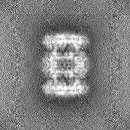
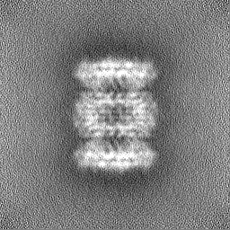
 Map data
 Sample
 Function and homology information
 proteasome endopeptidase complex / proteasome core complex, beta-subunit complex / proteasome core complex, alpha-subunit complex / threonine-type endopeptidase activity / proteasomal protein catabolic process / ubiquitin-dependent protein catabolic process /

 Authors
 Netherlands, 2 items
  Citation
 Structure visualization
 Downloads & links
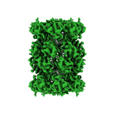 emd_12915.png
 http://ftp.pdbj.org/pub/emdb/structures/EMD-12915


 Z
Z Y
Y X
X


 Sample components
 Processing
 Electron microscopy
