 Movie
Movie Controller

[English] 日本語
 Yorodumi
- EMDB-12794: Cryo-EM structure of a twisted-dimer transthyretin amyloid fibril... -
+ Open data

- Basic information
| Entry | Database: EMDB / ID: EMD-12794 | |||||||||
|---|---|---|---|---|---|---|---|---|---|---|
| Title | Cryo-EM structure of a twisted-dimer transthyretin amyloid fibril from vitreous body of the eye | |||||||||
 Map data Map data | ||||||||||
 Sample Sample |
| |||||||||
| Function / homology |  Function and homology information Function and homology informationRetinoid cycle disease events /  thyroid hormone binding / The canonical retinoid cycle in rods (twilight vision) / Non-integrin membrane-ECM interactions / purine nucleobase metabolic process / Retinoid metabolism and transport / hormone activity / azurophil granule lumen / Amyloid fiber formation / Neutrophil degranulation ...Retinoid cycle disease events / thyroid hormone binding / The canonical retinoid cycle in rods (twilight vision) / Non-integrin membrane-ECM interactions / purine nucleobase metabolic process / Retinoid metabolism and transport / hormone activity / azurophil granule lumen / Amyloid fiber formation / Neutrophil degranulation / extracellular space / extracellular exosome / extracellular region / identical protein binding thyroid hormone binding / The canonical retinoid cycle in rods (twilight vision) / Non-integrin membrane-ECM interactions / purine nucleobase metabolic process / Retinoid metabolism and transport / hormone activity / azurophil granule lumen / Amyloid fiber formation / Neutrophil degranulation ...Retinoid cycle disease events / thyroid hormone binding / The canonical retinoid cycle in rods (twilight vision) / Non-integrin membrane-ECM interactions / purine nucleobase metabolic process / Retinoid metabolism and transport / hormone activity / azurophil granule lumen / Amyloid fiber formation / Neutrophil degranulation / extracellular space / extracellular exosome / extracellular region / identical protein bindingSimilarity search - Function | |||||||||
| Biological species |  Homo sapiens (human) / Human (human) Homo sapiens (human) / Human (human) | |||||||||
| Method | helical reconstruction / cryo EM / Resolution: 3.22 Å | |||||||||
 Authors Authors | Iakovleva I / Sauer-Eriksson AE | |||||||||
| Funding support |  Sweden, 1 items Sweden, 1 items
| |||||||||
 Citation Citation | Journal: Nat Commun / Year: 2021 Title: Structural basis for transthyretin amyloid formation in vitreous body of the eye. Authors: Irina Iakovleva / Michael Hall / Melanie Oelker / Linda Sandblad / Intissar Anan / A Elisabeth Sauer-Eriksson / Abstract: Amyloid transthyretin (ATTR) amyloidosis is characterized by the abnormal accumulation of ATTR fibrils in multiple organs. However, the structure of ATTR fibrils from the eye is poorly understood. ...Amyloid transthyretin (ATTR) amyloidosis is characterized by the abnormal accumulation of ATTR fibrils in multiple organs. However, the structure of ATTR fibrils from the eye is poorly understood. Here, we used cryo-EM to structurally characterize vitreous body ATTR fibrils. These structures were distinct from previously characterized heart fibrils, even though both have the same mutation and type A pathology. Differences were observed at several structural levels: in both the number and arrangement of protofilaments, and the conformation of the protein fibril in each layer of protofilaments. Thus, our results show that ATTR protein structure and its assembly into protofilaments in the type A fibrils can vary between patients carrying the same mutation. By analyzing and matching the interfaces between the amino acids in the ATTR fibril with those in the natively folded TTR, we are able to propose a mechanism for the structural conversion of TTR into a fibrillar form. | |||||||||
| History |
|
- Structure visualization
| Movie |
Movie viewer |
|---|---|
| Structure viewer | EM map: SurfViewMolmilJmol/JSmol |
| Supplemental images |

- Downloads & links
-EMDB archive
| Map data | emd_12794.map.gz | 13 MB | EMDB map data format | |
|---|---|---|---|---|
| Header (meta data) | emd-12794-v30.xmlemd-12794.xml | 10 KB 10 KB | Display Display | EMDB header |
| FSC (resolution estimation) | emd_12794_fsc.xml | 7.9 KB | Display | FSC data file |
| Images | 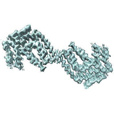 emd_12794.png emd_12794.png | 117.9 KB | ||
| Archive directory |  http://ftp.pdbj.org/pub/emdb/structures/EMD-12794ftp://ftp.pdbj.org/pub/emdb/structures/EMD-12794 http://ftp.pdbj.org/pub/emdb/structures/EMD-12794ftp://ftp.pdbj.org/pub/emdb/structures/EMD-12794 | HTTPS FTP |
-Related structure data
| Related structure data |  7ob4MC M: atomic model generated by this map C: citing same article ( |
|---|---|
| Similar structure data |


-Links
| EMDB pages | EMDB (EBI/PDBe) / EMDataResource |
|---|---|
| Related items in Molecule of the Month |


-Map
| File | Download / File: emd_12794.map.gz / Format: CCP4 / Size: 40.6 MB / Type: IMAGE STORED AS FLOATING POINT NUMBER (4 BYTES) | ||||||||||||||||||||||||||||||||||||||||||||||||||||||||||||
|---|---|---|---|---|---|---|---|---|---|---|---|---|---|---|---|---|---|---|---|---|---|---|---|---|---|---|---|---|---|---|---|---|---|---|---|---|---|---|---|---|---|---|---|---|---|---|---|---|---|---|---|---|---|---|---|---|---|---|---|---|---|
| Voxel size | X=Y=Z: 1.041 Å | ||||||||||||||||||||||||||||||||||||||||||||||||||||||||||||
| Density |
| ||||||||||||||||||||||||||||||||||||||||||||||||||||||||||||
| Symmetry | Space group: 1 | ||||||||||||||||||||||||||||||||||||||||||||||||||||||||||||
| Details | EMDB XML:
CCP4 map header:
| ||||||||||||||||||||||||||||||||||||||||||||||||||||||||||||
-Supplemental data
- Sample components
-Entire : Transthyretin derived amyloid fibril (V30M) from vitreous body
| Entire | Name: Transthyretin derived amyloid fibril (V30M) from vitreous body |
|---|---|
| Components |
|
-Supramolecule #1: Transthyretin derived amyloid fibril (V30M) from vitreous body
| Supramolecule | Name: Transthyretin derived amyloid fibril (V30M) from vitreous body type: tissue / ID: 1 / Parent: 0 / Macromolecule list: all |
|---|---|
| Source (natural) | Organism: Homo sapiens (human) / Organ: Vitreous body of the eye |
-Macromolecule #1: Transthyretin
| Macromolecule | Name: Transthyretin / type: protein_or_peptide / ID: 1 / Number of copies: 14 / Enantiomer: LEVO |
|---|---|
| Source (natural) | Organism: Human (human) |
| Molecular weight | Theoretical: 13.809426 KDa |
| Sequence | String: GPTGTGESKC PLMVKVLDAV RGSPAINVAM HVFRKAADDT WEPFASGKTS ESGELHGLTT EEEFVEGIYK VEIDTKSYWK ALGISPFHE HAEVVFTAND SGPRRYTIAA LLSPYSYSTT AVVTNPKE |
-Experimental details
-Structure determination
| Method | cryo EM |
|---|---|
 Processing Processing | helical reconstruction |
| Aggregation state | helical array |
-Sample preparation
| Buffer | pH: 7.4 |
|---|---|
| Vitrification | Cryogen name: ETHANE / Instrument: FEI VITROBOT MARK IV |
- Electron microscopy
| Microscope | FEI TITAN KRIOS |
|---|---|
| Electron beam | Acceleration voltage: 300 kV / Electron source: FIELD EMISSION GUN |
| Electron optics | Illumination mode: FLOOD BEAM / Imaging mode: BRIGHT FIELDBright-field microscopy |
| Image recording | Film or detector model: GATAN K2 SUMMIT (4k x 4k) / Average electron dose: 28.4 e/Å2 |
| Experimental equipment |  Model: Titan Krios / Image courtesy: FEI Company |
-Image processing
| Segment selection | Number selected: 130212 |
|---|---|
| Final angle assignment | Type: NOT APPLICABLE / Software - Name: RELION (ver. 3.1) |
| Final reconstruction | Applied symmetry - Helical parameters - Δz: 4.72 Å Applied symmetry - Helical parameters - Δ&Phi: -0.552 ° Applied symmetry - Helical parameters - Axial symmetry: C2 (2 fold cyclic )Resolution.type: BY AUTHOR / Resolution: 3.22 Å / Resolution method: FSC 0.143 CUT-OFF / Software - Name: RELION (ver. 3.1) / Number images used: 27778 |
| FSC plot (resolution estimation) |  |

