 Movie
Movie Controller

[English] 日本語
 Yorodumi
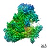
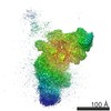
- PDB-6zfb: Structure of the B. subtilis RNA POLYMERASE in complex with HelD ... -
+ Open data

- Basic information
| Entry | Database: PDB / ID: 6zfb | |||||||||||||||||||||
|---|---|---|---|---|---|---|---|---|---|---|---|---|---|---|---|---|---|---|---|---|---|---|
| Title | Structure of the B. subtilis RNA POLYMERASE in complex with HelD (dimer) | |||||||||||||||||||||
 Components Components |
| |||||||||||||||||||||
 Keywords Keywords |  TRANSCRIPTION / TRANSCRIPTION/DNA/RNA / DNA-DEPENDENT RNA POLYMERASE / BACTERIAL / Helicase TRANSCRIPTION / TRANSCRIPTION/DNA/RNA / DNA-DEPENDENT RNA POLYMERASE / BACTERIAL / Helicase | |||||||||||||||||||||
| Function / homology |  Function and homology informationnucleoid / recombinational repair / 3'-5' DNA helicase activity / DNA helicase activity / DNA-directed RNA polymerase complex / ribonucleoside binding / DNA-directed 5'-3' RNA polymerase activity / DNA-directed RNA polymerase / DNA helicase / protein dimerization activity ...nucleoid / recombinational repair / 3'-5' DNA helicase activity / DNA helicase activity / DNA-directed RNA polymerase complex / ribonucleoside binding / DNA-directed 5'-3' RNA polymerase activity / DNA-directed RNA polymerase / DNA helicase / protein dimerization activity / hydrolase activity / response to antibiotic / DNA-templated transcription / regulation of DNA-templated transcription / magnesium ion binding / ATP hydrolysis activity / DNA binding / zinc ion binding / ATP binding / cytosol / cytoplasm Function and homology informationnucleoid / recombinational repair / 3'-5' DNA helicase activity / DNA helicase activity / DNA-directed RNA polymerase complex / ribonucleoside binding / DNA-directed 5'-3' RNA polymerase activity / DNA-directed RNA polymerase / DNA helicase / protein dimerization activity ...nucleoid / recombinational repair / 3'-5' DNA helicase activity / DNA helicase activity / DNA-directed RNA polymerase complex / ribonucleoside binding / DNA-directed 5'-3' RNA polymerase activity / DNA-directed RNA polymerase / DNA helicase / protein dimerization activity / hydrolase activity / response to antibiotic / DNA-templated transcription / regulation of DNA-templated transcription / magnesium ion binding / ATP hydrolysis activity / DNA binding / zinc ion binding / ATP binding / cytosol / cytoplasmSimilarity search - Function | |||||||||||||||||||||
| Biological species |  Bacillus subtilis (bacteria) Bacillus subtilis (bacteria) | |||||||||||||||||||||
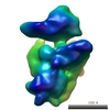
| Method | ELECTRON MICROSCOPY / single particle reconstruction / cryo EM / Resolution: 3.9 Å | |||||||||||||||||||||
 Authors Authors | Pei, H.-P. / Hilal, T. / Huang, Y.-H. / Said, N. / Loll, B. / Wahl, M.C. | |||||||||||||||||||||
| Funding support |  Germany, Germany,  United States, 6items United States, 6items
| |||||||||||||||||||||
 Citation Citation | Journal: Nat Commun / Year: 2020 Title: The δ subunit and NTPase HelD institute a two-pronged mechanism for RNA polymerase recycling. Authors: Hao-Hong Pei / Tarek Hilal / Zhuo A Chen / Yong-Heng Huang / Yuan Gao / Nelly Said / Bernhard Loll / Juri Rappsilber / Georgiy A Belogurov / Irina Artsimovitch / Markus C Wahl /   Abstract: Cellular RNA polymerases (RNAPs) can become trapped on DNA or RNA, threatening genome stability and limiting free enzyme pools, but how RNAP recycling into active states is achieved remains elusive. ...Cellular RNA polymerases (RNAPs) can become trapped on DNA or RNA, threatening genome stability and limiting free enzyme pools, but how RNAP recycling into active states is achieved remains elusive. In Bacillus subtilis, the RNAP δ subunit and NTPase HelD have been implicated in RNAP recycling. We structurally analyzed Bacillus subtilis RNAP-δ-HelD complexes. HelD has two long arms: a Gre cleavage factor-like coiled-coil inserts deep into the RNAP secondary channel, dismantling the active site and displacing RNA, while a unique helical protrusion inserts into the main channel, prying the β and β' subunits apart and, aided by δ, dislodging DNA. RNAP is recycled when, after releasing trapped nucleic acids, HelD dissociates from the enzyme in an ATP-dependent manner. HelD abundance during slow growth and a dimeric (RNAP-δ-HelD) structure that resembles hibernating eukaryotic RNAP I suggest that HelD might also modulate active enzyme pools in response to cellular cues. | |||||||||||||||||||||
| History |
|
- Structure visualization
| Movie |
Movie viewer |
|---|---|
| Structure viewer | Molecule: MolmilJmol/JSmol |
- Downloads & links
-Download
| PDBx/mmCIF format | 6zfb.cif.gz | 1.3 MB | Display | PDBx/mmCIF format |
|---|---|---|---|---|
| PDB format | pdb6zfb.ent.gz | 1 MB | Display | PDB format |
| PDBx/mmJSON format | 6zfb.json.gz | Tree view | PDBx/mmJSON format | |
| Others |  Other downloads Other downloads |
-Validation report
| Arichive directory | https://data.pdbj.org/pub/pdb/validation_reports/zf/6zfbftp://data.pdbj.org/pub/pdb/validation_reports/zf/6zfb | HTTPS FTP |
|---|
-Related structure data
| Related structure data | 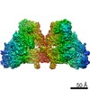 11105MC  6zcaC M: map data used to model this data C: citing same article ( |
|---|---|
| Similar structure data |


-Links
 PDBj
PDBj


- Assembly
| Deposited unit | 
|
|---|---|
| 1 |
|
-Components
-DNA-directed RNA polymerase subunit ... , 4 types, 10 molecules DdUVuvXxYy
| #1: Protein | Polymerase Mass: 16359.910 Da / Num. of mol.: 2 / Source method: isolated from a natural source / Source: (natural) Bacillus subtilis (bacteria) / References: UniProt: P12464*PLUS#3: Protein | Polymerase / RNAP subunit alpha / RNA polymerase subunit alpha / Transcriptase subunit alphaMass: 34842.387 Da / Num. of mol.: 4 / Source method: isolated from a natural source / Source: (natural) Bacillus subtilis (bacteria)References: UniProt: A0A063XB83, UniProt: P20429*PLUS, DNA-directed RNA polymerase#4: Protein | Polymerase / RNAP subunit beta / RNA polymerase subunit beta / Transcriptase subunit betaMass: 133847.938 Da / Num. of mol.: 2 / Source method: isolated from a natural source Details: Due to limited quality of the electron density, we were only able to trace some of the protein main chain as poly-Ala Source: (natural) Bacillus subtilis (bacteria)References: UniProt: A0A2J0WBQ0, UniProt: P37870*PLUS, DNA-directed RNA polymerase#5: Protein | Polymerase / RNAP subunit beta' / RNA polymerase subunit beta' / Transcriptase subunit beta'Mass: 134444.953 Da / Num. of mol.: 2 / Source method: isolated from a natural source / Source: (natural) Bacillus subtilis (bacteria)References: UniProt: A0A063XB23, UniProt: P37871*PLUS, DNA-directed RNA polymerase |
|---|
-Protein , 2 types, 4 molecules EeHh
| #2: Protein | Mass: 8228.357 Da / Num. of mol.: 2 / Source method: isolated from a natural source / Source: (natural) Bacillus subtilis (bacteria) / References: UniProt: A0A410WI33, UniProt: O31718*PLUS#6: Protein | HelicaseMass: 90052.516 Da / Num. of mol.: 2 / Source method: isolated from a natural source / Source: (natural) Bacillus subtilis (bacteria)References: UniProt: A0A164TSE8, UniProt: O32215*PLUS, DNA helicase |
|---|
-Non-polymers , 1 types, 2 molecules 
| #7: Chemical |  Mass: 65.409 Da / Num. of mol.: 2 / Source method: obtained synthetically / Formula: Zn Mass: 65.409 Da / Num. of mol.: 2 / Source method: obtained synthetically / Formula: Zn |
|---|
-Details
| Has ligand of interest | N |
|---|
-Experimental details
-Experiment
| Experiment | Method: ELECTRON MICROSCOPY |
|---|---|
| EM experiment | Aggregation state: PARTICLE / 3D reconstruction method: single particle reconstruction |
- Sample preparation
| Component | Name: RNA POLYMERASE in complex with HelD / Type: COMPLEX / Entity ID: #1-#6 / Source: NATURAL |
|---|---|
| Molecular weight | Value: 0.9 MDa / Experimental value: NO |
| Source (natural) | Organism: Bacillus subtilis (bacteria) |
| Buffer solution | pH: 7.6 |
| Specimen | Conc.: 5 mg/ml / Embedding applied: NO / Shadowing applied: NO / Staining applied: NO / Vitrification applied: YES |
| Vitrification | Instrument: FEI VITROBOT MARK IV / Cryogen name: ETHANE / Humidity: 100 % / Chamber temperature: 283 K |
- Electron microscopy imaging
| Experimental equipment |  Model: Titan Krios / Image courtesy: FEI Company |
|---|---|
| Microscopy | Model: FEI TITAN KRIOS |
| Electron gun | Electron source: FIELD EMISSION GUN / Accelerating voltage: 300 kV / Illumination mode: FLOOD BEAM |
| Electron lens | Mode: BRIGHT FIELDBright-field microscopy / Nominal magnification: 96000 X / Nominal defocus max: 2500 nm / Nominal defocus min: 1000 nm / Cs: 2.7 mm / C2 aperture diameter: 50 µm / Alignment procedure: ZEMLIN TABLEAU |
| Specimen holder | Specimen holder model: FEI TITAN KRIOS AUTOGRID HOLDER |
| Image recording | Electron dose: 40 e/Å2 / Detector mode: COUNTING / Film or detector model: FEI FALCON III (4k x 4k) |
- Processing
| Software |
| ||||||||||||||||||||||||||||
|---|---|---|---|---|---|---|---|---|---|---|---|---|---|---|---|---|---|---|---|---|---|---|---|---|---|---|---|---|---|
| EM software |
| ||||||||||||||||||||||||||||
| CTF correction | Type: PHASE FLIPPING AND AMPLITUDE CORRECTION | ||||||||||||||||||||||||||||
| Symmetry | Point symmetry: C1 (asymmetric) | ||||||||||||||||||||||||||||
| 3D reconstruction | Resolution: 3.9 Å / Resolution method: FSC 0.143 CUT-OFF / Num. of particles: 176374 / Symmetry type: POINT | ||||||||||||||||||||||||||||
| Refinement | Cross valid method: NONE Stereochemistry target values: GeoStd + Monomer Library + CDL v1.2 | ||||||||||||||||||||||||||||
| Displacement parameters | Biso mean: 95.58 Å2 | ||||||||||||||||||||||||||||
| Refine LS restraints |
|
