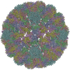
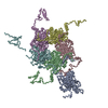
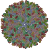
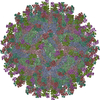
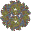
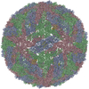
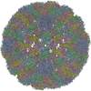
CryoEM Structure of Merkel Cell Polyomavirus Virus-like Particle
要素
Capsid protein VP1カプシド
キーワード
VIRUS LIKE PARTICLE (ウイルス様粒子) / polyomavirus (ポリオーマウイルス科) / capsid (カプシド) / Merkel cell carcinoma / jelly roll fold
機能・相同性
Capsid protein VP1,Polyomavirus / Polyomavirus capsid protein VP1 superfamily / Polyomavirus coat protein / Double-stranded DNA virus, group I, capsid / カプシド / structural molecule activity / Capsid protein VP1
ジャーナル: J Virol / 年: 2020 タイトル: Structure of Merkel Cell Polyomavirus Capsid and Interaction with Its Glycosaminoglycan Attachment Receptor. 著者: Niklas J Bayer / Dovile Januliene / Georg Zocher / Thilo Stehle / Arne Moeller / Bärbel S Blaum / 要旨: Merkel cell polyomavirus (MCPyV) is a human double-stranded DNA tumor virus. MCPyV cell entry is unique among members of the polyomavirus family as it requires the engagement of two types of glycans, ...Merkel cell polyomavirus (MCPyV) is a human double-stranded DNA tumor virus. MCPyV cell entry is unique among members of the polyomavirus family as it requires the engagement of two types of glycans, sialylated oligosaccharides and sulfated glycosaminoglycans (GAGs). Here, we present crystallographic and cryo-electron microscopic structures of the icosahedral MCPyV capsid and analysis of its glycan interactions via nuclear magnetic resonance (NMR) spectroscopy. While sialic acid binding is specific for α2-3-linked sialic acid and mediated by the exposed apical loops of the major capsid protein VP1, a broad range of GAG oligosaccharides bind to recessed regions between VP1 capsomers. Individual VP1 capsomers are tethered to one another by an extensive disulfide network that differs in architecture from previously described interactions for other PyVs. An unusual C-terminal extension in MCPyV VP1 projects from the recessed capsid regions. Mutagenesis experiments show that this extension is dispensable for receptor interactions. The MCPyV genome was found to be clonally integrated in 80% of cases of Merkel cell carcinoma (MCC), a rare but aggressive form of human skin cancer, strongly suggesting that this virus is tumorigenic. In the metastasizing state, the course of the disease is often fatal, especially in immunocompromised individuals, as reflected by the high mortality rate of 33 to 46% and the low 5-year survival rate (<45%). The high seroprevalence of about 60% makes MCPyV a serious health care burden and illustrates the need for targeted treatments. In this study, we present the first high-resolution structural data for this human tumor virus and demonstrate that the full capsid is required for the essential interaction with its GAG receptor(s). Together, these data can be used as a basis for future strategies in drug development.
 ムービー
ムービー コントローラー


 データを開く
 基本情報
基本情報 要素
要素 カプシド
カプシド  キーワード
キーワード 機能・相同性情報
 データ登録者
 ドイツ, 3件
  引用
引用
 構造の表示
構造の表示 ダウンロードとリンク
 その他のダウンロード
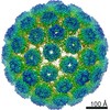


 PDBj
PDBj


 集合体
集合体

 試料調製
試料調製 電子顕微鏡撮影
 解析
解析