 Movie
Movie Controller

[English] 日本語
 Yorodumi
- EMDB-12534: Structure of the yeast Gcn1 bound to a leading stalled 80S riboso... -
+ Open data

- Basic information
| Entry | Database: EMDB / ID: EMD-12534 | |||||||||
|---|---|---|---|---|---|---|---|---|---|---|
| Title | Structure of the yeast Gcn1 bound to a leading stalled 80S ribosome with Rbg2, Gir2, A- and P-tRNA and eIF5A | |||||||||
 Map data Map data | ||||||||||
 Sample Sample |
| |||||||||
| Function / homology |  Function and homology information Function and homology informationpositive regulation of cytoplasmic translational elongation through polyproline stretches / Hypusine synthesis from eIF5A-lysine / CAT tailing /  translational frameshifting / positive regulation of translational termination / positive regulation of translational elongation / maturation of SSU-rRNA from tricistronic rRNA transcript (SSU-rRNA, LSU-rRNA,5S) / positive regulation of cellular response to amino acid starvation / negative regulation of glucose mediated signaling pathway / negative regulation of translational frameshifting ...positive regulation of cytoplasmic translational elongation through polyproline stretches / Hypusine synthesis from eIF5A-lysine / CAT tailing / translational frameshifting / positive regulation of translational termination / positive regulation of translational elongation / maturation of SSU-rRNA from tricistronic rRNA transcript (SSU-rRNA, LSU-rRNA,5S) / positive regulation of cellular response to amino acid starvation / negative regulation of glucose mediated signaling pathway / negative regulation of translational frameshifting / Negative regulators of DDX58/IFIH1 signaling / Protein methylation / : / positive regulation of translational fidelity / RMTs methylate histone arginines / mTORC1-mediated signalling / ribosome-associated ubiquitin-dependent protein catabolic process / Protein hydroxylation / GDP-dissociation inhibitor activity / pre-mRNA 5'-splice site binding / positive regulation of nuclear-transcribed mRNA catabolic process, deadenylation-dependent decay / Formation of the ternary complex, and subsequently, the 43S complex / Translation initiation complex formation / cleavage in ITS2 between 5.8S rRNA and LSU-rRNA of tricistronic rRNA transcript (SSU-rRNA, 5.8S rRNA, LSU-rRNA) / Ribosomal scanning and start codon recognition / preribosome, small subunit precursor / translational elongation / response to cycloheximide / mRNA destabilization / Major pathway of rRNA processing in the nucleolus and cytosol / SRP-dependent cotranslational protein targeting to membrane / GTP hydrolysis and joining of the 60S ribosomal subunit / Formation of a pool of free 40S subunits / Nonsense Mediated Decay (NMD) independent of the Exon Junction Complex (EJC) / Nonsense Mediated Decay (NMD) enhanced by the Exon Junction Complex (EJC) / endonucleolytic cleavage to generate mature 3'-end of SSU-rRNA from (SSU-rRNA, 5.8S rRNA, LSU-rRNA) / negative regulation of mRNA splicing, via spliceosome / ribosomal small subunit export from nucleus / positive regulation of translational initiation / preribosome, large subunit precursor / L13a-mediated translational silencing of Ceruloplasmin expression / translation regulator activity / 90S preribosome / protein-RNA complex assembly / ribosomal large subunit export from nucleus / Ub-specific processing proteases / G-protein alpha-subunit binding / regulation of translational fidelity / positive regulation of protein kinase activity / endonucleolytic cleavage in ITS1 to separate SSU-rRNA from 5.8S rRNA and LSU-rRNA from tricistronic rRNA transcript (SSU-rRNA, 5.8S rRNA, LSU-rRNA) / ribosomal subunit export from nucleus / translation elongation factor activity / translational termination / rescue of stalled ribosome / maturation of LSU-rRNA from tricistronic rRNA transcript (SSU-rRNA, 5.8S rRNA, LSU-rRNA) / maturation of SSU-rRNA from tricistronic rRNA transcript (SSU-rRNA, 5.8S rRNA, LSU-rRNA) / maturation of SSU-rRNA / ribosomal large subunit biogenesis / DNA-(apurinic or apyrimidinic site) endonuclease activity / maturation of LSU-rRNA / cellular response to amino acid starvation / translation initiation factor activity / ribosome assembly / small-subunit processome / protein kinase C binding / maintenance of translational fidelity / macroautophagy / modification-dependent protein catabolic process / ribosomal large subunit assembly / rRNA processing / ribosomal small subunit biogenesis / protein tag activity / small ribosomal subunit rRNA binding / cytoplasmic stress granule / ribosomal small subunit assembly / ribosome binding / ribosome biogenesis / large ribosomal subunit rRNA binding / small ribosomal subunit / cytosolic small ribosomal subunit / 5S rRNA binding / cytoplasmic translation / cytosolic large ribosomal subunit / negative regulation of translation / rRNA binding / protein ubiquitination / ribosome / structural constituent of ribosome / positive regulation of protein phosphorylation / translation / G protein-coupled receptor signaling pathway / response to antibiotic / negative regulation of gene expression / mRNA binding / ubiquitin protein ligase binding / GTP binding / nucleolus / perinuclear region of cytoplasm / mitochondrion / RNA binding translational frameshifting / positive regulation of translational termination / positive regulation of translational elongation / maturation of SSU-rRNA from tricistronic rRNA transcript (SSU-rRNA, LSU-rRNA,5S) / positive regulation of cellular response to amino acid starvation / negative regulation of glucose mediated signaling pathway / negative regulation of translational frameshifting ...positive regulation of cytoplasmic translational elongation through polyproline stretches / Hypusine synthesis from eIF5A-lysine / CAT tailing / translational frameshifting / positive regulation of translational termination / positive regulation of translational elongation / maturation of SSU-rRNA from tricistronic rRNA transcript (SSU-rRNA, LSU-rRNA,5S) / positive regulation of cellular response to amino acid starvation / negative regulation of glucose mediated signaling pathway / negative regulation of translational frameshifting / Negative regulators of DDX58/IFIH1 signaling / Protein methylation / : / positive regulation of translational fidelity / RMTs methylate histone arginines / mTORC1-mediated signalling / ribosome-associated ubiquitin-dependent protein catabolic process / Protein hydroxylation / GDP-dissociation inhibitor activity / pre-mRNA 5'-splice site binding / positive regulation of nuclear-transcribed mRNA catabolic process, deadenylation-dependent decay / Formation of the ternary complex, and subsequently, the 43S complex / Translation initiation complex formation / cleavage in ITS2 between 5.8S rRNA and LSU-rRNA of tricistronic rRNA transcript (SSU-rRNA, 5.8S rRNA, LSU-rRNA) / Ribosomal scanning and start codon recognition / preribosome, small subunit precursor / translational elongation / response to cycloheximide / mRNA destabilization / Major pathway of rRNA processing in the nucleolus and cytosol / SRP-dependent cotranslational protein targeting to membrane / GTP hydrolysis and joining of the 60S ribosomal subunit / Formation of a pool of free 40S subunits / Nonsense Mediated Decay (NMD) independent of the Exon Junction Complex (EJC) / Nonsense Mediated Decay (NMD) enhanced by the Exon Junction Complex (EJC) / endonucleolytic cleavage to generate mature 3'-end of SSU-rRNA from (SSU-rRNA, 5.8S rRNA, LSU-rRNA) / negative regulation of mRNA splicing, via spliceosome / ribosomal small subunit export from nucleus / positive regulation of translational initiation / preribosome, large subunit precursor / L13a-mediated translational silencing of Ceruloplasmin expression / translation regulator activity / 90S preribosome / protein-RNA complex assembly / ribosomal large subunit export from nucleus / Ub-specific processing proteases / G-protein alpha-subunit binding / regulation of translational fidelity / positive regulation of protein kinase activity / endonucleolytic cleavage in ITS1 to separate SSU-rRNA from 5.8S rRNA and LSU-rRNA from tricistronic rRNA transcript (SSU-rRNA, 5.8S rRNA, LSU-rRNA) / ribosomal subunit export from nucleus / translation elongation factor activity / translational termination / rescue of stalled ribosome / maturation of LSU-rRNA from tricistronic rRNA transcript (SSU-rRNA, 5.8S rRNA, LSU-rRNA) / maturation of SSU-rRNA from tricistronic rRNA transcript (SSU-rRNA, 5.8S rRNA, LSU-rRNA) / maturation of SSU-rRNA / ribosomal large subunit biogenesis / DNA-(apurinic or apyrimidinic site) endonuclease activity / maturation of LSU-rRNA / cellular response to amino acid starvation / translation initiation factor activity / ribosome assembly / small-subunit processome / protein kinase C binding / maintenance of translational fidelity / macroautophagy / modification-dependent protein catabolic process / ribosomal large subunit assembly / rRNA processing / ribosomal small subunit biogenesis / protein tag activity / small ribosomal subunit rRNA binding / cytoplasmic stress granule / ribosomal small subunit assembly / ribosome binding / ribosome biogenesis / large ribosomal subunit rRNA binding / small ribosomal subunit / cytosolic small ribosomal subunit / 5S rRNA binding / cytoplasmic translation / cytosolic large ribosomal subunit / negative regulation of translation / rRNA binding / protein ubiquitination / ribosome / structural constituent of ribosome / positive regulation of protein phosphorylation / translation / G protein-coupled receptor signaling pathway / response to antibiotic / negative regulation of gene expression / mRNA binding / ubiquitin protein ligase binding / GTP binding / nucleolus / perinuclear region of cytoplasm / mitochondrion / RNA bindingSimilarity search - Function | |||||||||
| Biological species |  Saccharomyces cerevisiae S288C (yeast) Saccharomyces cerevisiae S288C (yeast) | |||||||||
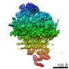
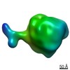
| Method | single particle reconstruction / cryo EM / Resolution: 3.9 Å | |||||||||
 Authors Authors | Pochopien AA / Beckert B / Wilson DN | |||||||||
| Funding support |  Germany, 2 items Germany, 2 items
| |||||||||
 Citation Citation | Journal: Proc Natl Acad Sci U S A / Year: 2021 Title: Structure of Gcn1 bound to stalled and colliding 80S ribosomes. Authors: Agnieszka A Pochopien / Bertrand Beckert / Sergo Kasvandik / Otto Berninghausen / Roland Beckmann / Tanel Tenson / Daniel N Wilson /  Abstract: The Gcn pathway is conserved in all eukaryotes, including mammals such as humans, where it is a crucial part of the integrated stress response (ISR). Gcn1 serves as an essential effector protein for ...The Gcn pathway is conserved in all eukaryotes, including mammals such as humans, where it is a crucial part of the integrated stress response (ISR). Gcn1 serves as an essential effector protein for the kinase Gcn2, which in turn is activated by stalled ribosomes, leading to phosphorylation of eIF2 and a subsequent global repression of translation. The fine-tuning of this adaptive response is performed by the Rbg2/Gir2 complex, a negative regulator of Gcn2. Despite the wealth of available biochemical data, information on structures of Gcn proteins on the ribosome has remained elusive. Here we present a cryo-electron microscopy structure of the yeast Gcn1 protein in complex with stalled and colliding 80S ribosomes. Gcn1 interacts with both 80S ribosomes within the disome, such that the Gcn1 HEAT repeats span from the P-stalk region on the colliding ribosome to the P-stalk and the A-site region of the lead ribosome. The lead ribosome is stalled in a nonrotated state with peptidyl-tRNA in the A-site, uncharged tRNA in the P-site, eIF5A in the E-site, and Rbg2/Gir2 in the A-site factor binding region. By contrast, the colliding ribosome adopts a rotated state with peptidyl-tRNA in a hybrid A/P-site, uncharged-tRNA in the P/E-site, and Mbf1 bound adjacent to the mRNA entry channel on the 40S subunit. Collectively, our findings reveal the interaction mode of the Gcn2-activating protein Gcn1 with colliding ribosomes and provide insight into the regulation of Gcn2 activation. The binding of Gcn1 to a disome has important implications not only for the Gcn2-activated ISR, but also for the general ribosome-associated quality control pathways. | |||||||||
| History |
|
- Structure visualization
| Movie |
Movie viewer |
|---|---|
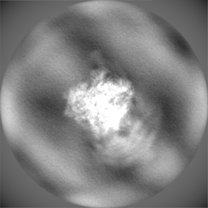
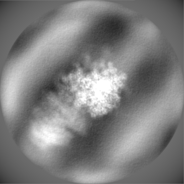
| Structure viewer | EM map: SurfViewMolmilJmol/JSmol |
| Supplemental images |

- Downloads & links
-EMDB archive
| Map data | emd_12534.map.gz | 689.6 MB | EMDB map data format | |
|---|---|---|---|---|
| Header (meta data) | emd-12534-v30.xmlemd-12534.xml | 113.4 KB 113.4 KB | Display Display | EMDB header |
| FSC (resolution estimation) | emd_12534_fsc.xml | 24.8 KB | Display | FSC data file |
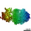
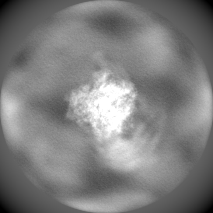
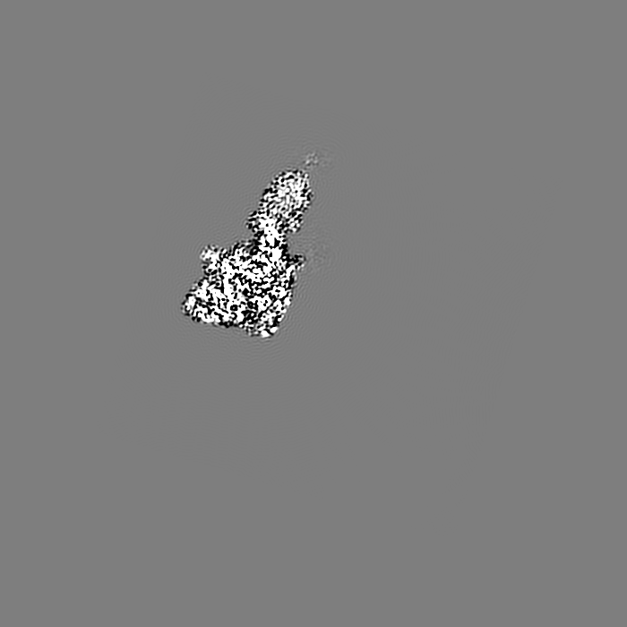
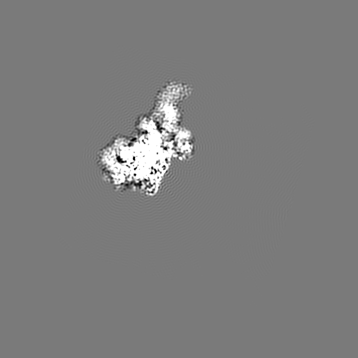
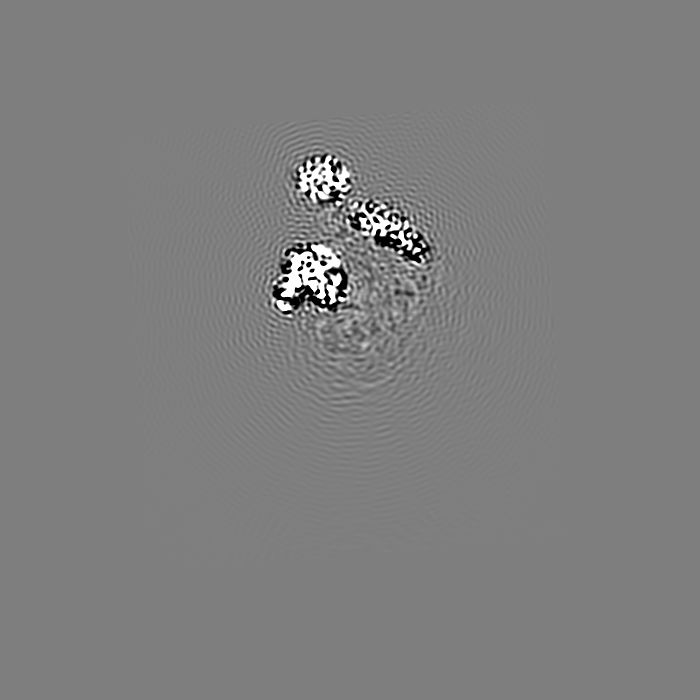
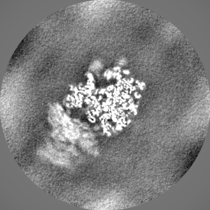
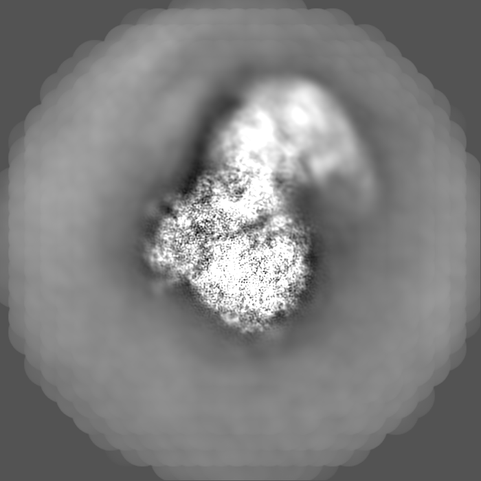
| Images | 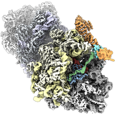 emd_12534.png emd_12534.png | 248 KB | ||
| Others | emd_12534_additional_1.map.gzemd_12534_additional_2.map.gzemd_12534_additional_3.map.gzemd_12534_additional_4.map.gzemd_12534_additional_5.map.gzemd_12534_additional_6.map.gzemd_12534_additional_7.map.gzemd_12534_additional_8.map.gzemd_12534_half_map_1.map.gzemd_12534_half_map_2.map.gz | 265.7 MB 265.6 MB 1 GB 98.2 MB 265.8 MB 265.7 MB 1 GB 687.9 MB 1 GB 1 GB | ||
| Archive directory |  http://ftp.pdbj.org/pub/emdb/structures/EMD-12534ftp://ftp.pdbj.org/pub/emdb/structures/EMD-12534 http://ftp.pdbj.org/pub/emdb/structures/EMD-12534ftp://ftp.pdbj.org/pub/emdb/structures/EMD-12534 | HTTPS FTP |
-Related structure data
| Related structure data |  7nrcMC  7nrdC M: atomic model generated by this map C: citing same article ( |
|---|---|
| Similar structure data |


-Links
| EMDB pages | EMDB (EBI/PDBe) / EMDataResource |
|---|---|
| Related items in Molecule of the Month |


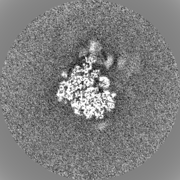
-Map
| File | Download / File: emd_12534.map.gz / Format: CCP4 / Size: 1.3 GB / Type: IMAGE STORED AS FLOATING POINT NUMBER (4 BYTES) | ||||||||||||||||||||||||||||||||||||||||||||||||||||||||||||
|---|---|---|---|---|---|---|---|---|---|---|---|---|---|---|---|---|---|---|---|---|---|---|---|---|---|---|---|---|---|---|---|---|---|---|---|---|---|---|---|---|---|---|---|---|---|---|---|---|---|---|---|---|---|---|---|---|---|---|---|---|---|
| Voxel size | X=Y=Z: 1.084 Å | ||||||||||||||||||||||||||||||||||||||||||||||||||||||||||||
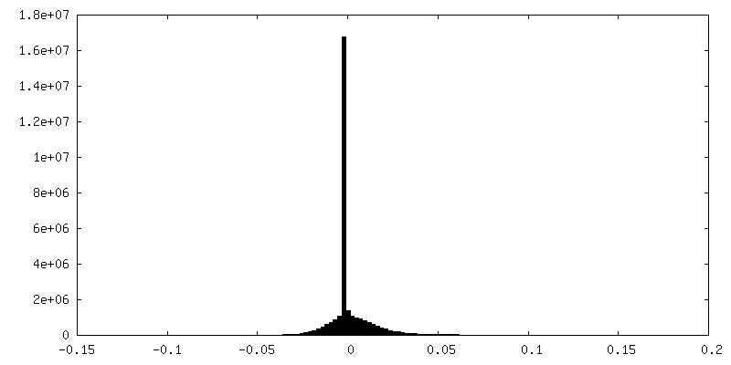
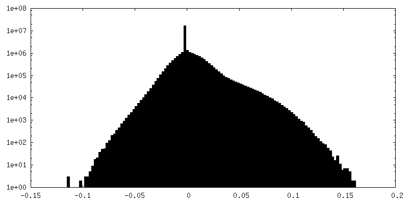
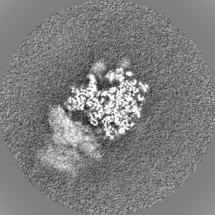
| Density |
| ||||||||||||||||||||||||||||||||||||||||||||||||||||||||||||
| Symmetry | Space group: 1 | ||||||||||||||||||||||||||||||||||||||||||||||||||||||||||||
| Details | EMDB XML:
CCP4 map header:
| ||||||||||||||||||||||||||||||||||||||||||||||||||||||||||||
-Supplemental data
+Additional map: #1
 Z
Z Y
Y X
X


+Additional map: #2


+Additional map: #3


+Additional map: #4


+Additional map: #5


+Additional map: #6


+Additional map: #7


+Additional map: #8


+Half map: #1


+Half map: #2


- Sample components
+Entire : Structure of the yeast Gcn1-bound leading stalled 80S ribosome wi...
+Supramolecule #1: Structure of the yeast Gcn1-bound leading stalled 80S ribosome wi...
+Macromolecule #1: 18S rRNA (1771-MER)
+Macromolecule #2: RNA (5'-R(P*AP*UP*GP*AP*AP*A)-3')
+Macromolecule #36: tRNA (76-MER)
+Macromolecule #37: tRNA (75-MER)
+Macromolecule #40: 25S rRNA (3184-MER)
+Macromolecule #41: 5S rRNA (121-MER)
+Macromolecule #42: 5.8S rRNA (158-MER)
+Macromolecule #3: 40S ribosomal protein S0-A
+Macromolecule #4: 40S ribosomal protein S1-A
+Macromolecule #5: 40S ribosomal protein S15
+Macromolecule #6: 40S ribosomal protein S2
+Macromolecule #7: 40S ribosomal protein S3
+Macromolecule #8: 40S ribosomal protein S4-A
+Macromolecule #9: 40S ribosomal protein S5
+Macromolecule #10: 40S ribosomal protein S6-A
+Macromolecule #11: 40S ribosomal protein S7-A
+Macromolecule #12: 40S ribosomal protein S8-A
+Macromolecule #13: 40S ribosomal protein S9-A
+Macromolecule #14: 40S ribosomal protein S10-A
+Macromolecule #15: 40S ribosomal protein S11-A
+Macromolecule #16: 40S ribosomal protein S12
+Macromolecule #17: 40S ribosomal protein S13
+Macromolecule #18: 40S ribosomal protein S14-B
+Macromolecule #19: 40S ribosomal protein S16-A
+Macromolecule #20: 40S ribosomal protein S17-B
+Macromolecule #21: 40S ribosomal protein S18-A
+Macromolecule #22: 40S ribosomal protein S19-A
+Macromolecule #23: 40S ribosomal protein S20
+Macromolecule #24: 40S ribosomal protein S21-A
+Macromolecule #25: 40S ribosomal protein S22-A
+Macromolecule #26: 40S ribosomal protein S23-A
+Macromolecule #27: 40S ribosomal protein S24-A
+Macromolecule #28: 40S ribosomal protein S25-A
+Macromolecule #29: 40S ribosomal protein S26-B
+Macromolecule #30: 40S ribosomal protein S27-A
+Macromolecule #31: 40S ribosomal protein S29-A
+Macromolecule #32: 40S ribosomal protein S30-A
+Macromolecule #33: 40S ribosomal protein S31
+Macromolecule #34: Guanine nucleotide-binding protein subunit beta-like protein
+Macromolecule #35: 40S ribosomal protein S28-A
+Macromolecule #38: Ribosome-interacting GTPase 2
+Macromolecule #39: GIR2
+Macromolecule #43: 60S ribosomal protein L2-A
+Macromolecule #44: 60S ribosomal protein L3
+Macromolecule #45: 60S ribosomal protein L4-A
+Macromolecule #46: 60S ribosomal protein L5
+Macromolecule #47: 60S ribosomal protein L6-B
+Macromolecule #48: 60S ribosomal protein L7-A
+Macromolecule #49: 60S ribosomal protein L8-A
+Macromolecule #50: 60S ribosomal protein L9-A
+Macromolecule #51: 60S ribosomal protein L10
+Macromolecule #52: 60S ribosomal protein L11-B
+Macromolecule #53: 60S ribosomal protein L13-A
+Macromolecule #54: 60S ribosomal protein L14-A
+Macromolecule #55: 60S ribosomal protein L15-A
+Macromolecule #56: 60S ribosomal protein L16-A
+Macromolecule #57: 60S ribosomal protein L17-A
+Macromolecule #58: 60S ribosomal protein L18-A
+Macromolecule #59: 60S ribosomal protein L19-A
+Macromolecule #60: 60S ribosomal protein L20-A
+Macromolecule #61: 60S ribosomal protein L21-A
+Macromolecule #62: 60S ribosomal protein L22-A
+Macromolecule #63: 60S ribosomal protein L23-A
+Macromolecule #64: 60S ribosomal protein L24-A
+Macromolecule #65: 60S ribosomal protein L25
+Macromolecule #66: 60S ribosomal protein L26-A
+Macromolecule #67: 60S ribosomal protein L27-A
+Macromolecule #68: 60S ribosomal protein L28
+Macromolecule #69: 60S ribosomal protein L29
+Macromolecule #70: 60S ribosomal protein L30
+Macromolecule #71: 60S ribosomal protein L31-A
+Macromolecule #72: 60S ribosomal protein L32
+Macromolecule #73: 60S ribosomal protein L33-A
+Macromolecule #74: 60S ribosomal protein L34-A
+Macromolecule #75: 60S ribosomal protein L35-A
+Macromolecule #76: 60S ribosomal protein L36-A
+Macromolecule #77: 60S ribosomal protein L37-A
+Macromolecule #78: 60S ribosomal protein L38
+Macromolecule #79: 60S ribosomal protein L39
+Macromolecule #80: 60S ribosomal protein L40-A
+Macromolecule #81: 60S ribosomal protein L41-A
+Macromolecule #82: 60S ribosomal protein L42-A
+Macromolecule #83: 60S ribosomal protein L43-A
+Macromolecule #84: eiF5A
+Macromolecule #85: L1 60S ribosomal protein
+Macromolecule #86: GCN1
-Experimental details
-Structure determination
| Method | cryo EM |
|---|---|
 Processing Processing | single particle reconstruction |
| Aggregation state | particle |
-Sample preparation
| Buffer | pH: 7.5 |
|---|---|
| Vitrification | Cryogen name: ETHANE-PROPANE |
- Electron microscopy
| Microscope | FEI TITAN KRIOS |
|---|---|
| Electron beam | Acceleration voltage: 300 kV / Electron source: FIELD EMISSION GUN |
| Electron optics | Illumination mode: SPOT SCAN / Imaging mode: BRIGHT FIELDBright-field microscopy |
| Image recording | Film or detector model: FEI FALCON II (4k x 4k) / Average electron dose: 2.5 e/Å2 |
| Experimental equipment |  Model: Titan Krios / Image courtesy: FEI Company |
-Image processing
| Initial angle assignment | Type: MAXIMUM LIKELIHOOD |
|---|---|
| Final angle assignment | Type: MAXIMUM LIKELIHOOD |
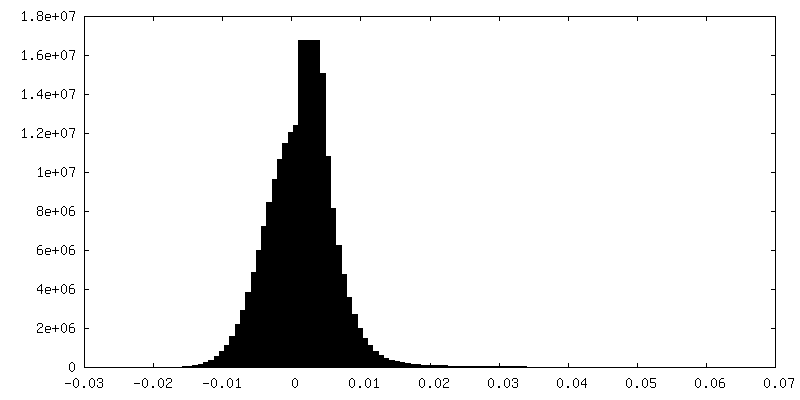
| Final reconstruction | Applied symmetry - Point group: C1 (asymmetric) / Resolution.type: BY AUTHOR / Resolution: 3.9 Å / Resolution method: FSC 0.143 CUT-OFF / Number images used: 30016 |
| FSC plot (resolution estimation) |  |
