Movie
Movie Controller
 Structure viewers
 About Yorodumi Papers


+Search query
-Structure paper
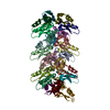
| Title | pH- and concentration-dependent supramolecular assembly of a fungal defensin plectasin variant into helical non-amyloid fibrils. |
|---|---|
| Journal, issue, pages | Nat Commun, Vol. 13, Issue 1, Page 3162, Year 2022 |
| Publish date | Jun 7, 2022 |
 Authors Authors | Christin Pohl / Gregory Effantin / Eaazhisai Kandiah / Sebastian Meier / Guanghong Zeng / Werner Streicher / Dorotea Raventos Segura / Per H Mygind / Dorthe Sandvang / Line Anker Nielsen / Günther H J Peters / Guy Schoehn / Christoph Mueller-Dieckmann / Allan Noergaard / Pernille Harris /     |
| PubMed Abstract | Self-assembly and fibril formation play important roles in protein behaviour. Amyloid fibril formation is well-studied due to its role in neurodegenerative diseases and characterized by refolding of ...Self-assembly and fibril formation play important roles in protein behaviour. Amyloid fibril formation is well-studied due to its role in neurodegenerative diseases and characterized by refolding of the protein into predominantly β-sheet form. However, much less is known about the assembly of proteins into other types of supramolecular structures. Using cryo-electron microscopy at a resolution of 1.97 Å, we show that a triple-mutant of the anti-microbial peptide plectasin, PPI42, assembles into helical non-amyloid fibrils. The in vitro anti-microbial activity was determined and shown to be enhanced compared to the wildtype. Plectasin contains a cysteine-stabilised α-helix-β-sheet structure, which remains intact upon fibril formation. Two protofilaments form a right-handed protein fibril. The fibril formation is reversible and follows sigmoidal kinetics with a pH- and concentration dependent equilibrium between soluble monomer and protein fibril. This high-resolution structure reveals that α/β proteins can natively assemble into fibrils. |
 External links External links | Nat Commun / PubMed:35672293 / PubMed Central |
| Methods | EM (helical sym.) / X-ray diffraction |
| Resolution | 1.131 - 3.4 Å |
| Structure data | EMDB-12775, PDB-7oae: EMDB-12776, PDB-7oag:  PDB-7o76: |
| Chemicals |  ChemComp-PEG:  ChemComp-HOH: |
| Source |
|
 Keywords Keywords |  ANTIMICROBIAL PROTEIN / fungal defensin / fibril / helical ANTIMICROBIAL PROTEIN / fungal defensin / fibril / helical |