ムービー
ムービー コントローラー
 構造ビューア
 万見文献について


+検索条件
-Structure paper
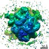
| タイトル | Cryo-EM Structures Reveal Relocalization of MetAP in the Presence of Other Protein Biogenesis Factors at the Ribosomal Tunnel Exit. |
|---|---|
| ジャーナル・号・ページ | J Mol Biol, Vol. 431, Issue 7, Page 1426-1439, Year 2019 |
| 掲載日 | 2019年3月29日 |
 著者 著者 | Sayan Bhakta / Shirin Akbar / Jayati Sengupta /  |
| PubMed 要旨 | During protein biosynthesis in bacteria, one of the earliest events that a nascent polypeptide chain goes through is the co-translational enzymatic processing. The event includes two enzymatic ...During protein biosynthesis in bacteria, one of the earliest events that a nascent polypeptide chain goes through is the co-translational enzymatic processing. The event includes two enzymatic pathways: deformylation of the N-terminal methionine by the enzyme peptide deformylase (PDF), followed by methionine excision catalyzed by methionine aminopeptidase (MetAP). During the enzymatic processing, the emerging nascent protein likely remains shielded by the ribosome-associated chaperone trigger factor. The ribosome tunnel exit serves as a stage for recruiting proteins involved in maturation processes of the nascent chain. Co-translational processing of nascent chains is a critical step for subsequent folding and functioning of mature proteins. Here, we present cryo-electron microscopy structures of Escherichia coli (E. coli) ribosome in complex with the nascent chain processing proteins. The structures reveal overlapping binding sites for PDF and MetAP when they bind individually at the tunnel exit site, where L22-L32 protein region provides primary anchoring sites for both proteins. In the absence of PDF, trigger factor can access ribosomal tunnel exit when MetAP occupies its primary binding site. Interestingly, however, in the presence of PDF, when MetAP's primary binding site is already engaged, MetAP has a remarkable ability to occupy an alternative binding site adjacent to PDF. Our study, thus, discloses an unexpected mechanism that MetAP adopts for context-specific ribosome association. |
 リンク リンク | J Mol Biol / PubMed:30753870 |
| 手法 | EM (単粒子) |
| 解像度 | 10.5 - 14.2 Å |
| 構造データ | EMDB-9750: Cryo-EM density map of E. coli 70S ribosome in complex with peptide deformylase enzyme EMDB-9752: Cryo-EM density map of E. coli 70S ribosome in complex with methionine aminopeptidase enzyme EMDB-9753: Cryo-EM density map of peptide deformylase and methionine aminopeptidase bound to the E. coli 70S ribosome EMDB-9759: Cryo-EM density map of methionine aminopeptidase enzyme and chaperone trigger factor bound to the E. coli 70S ribosome |
| 由来 |
|
 キーワード キーワード |  RIBOSOME (リボソーム) / E. coli 70S ribosome / pepdite deformylase / Nascent polypeptide exit tunnel / Protein biogenesis / Methionine aminopeptidase (メチオニルアミノペプチダーゼ) / Methionine excision / Polypeptide exit tunnel / Peptide deformylase (ペプチドデホルミラーゼ) / Chaperone / Trigger factor / PPIase (プロリルイソメラーゼ) RIBOSOME (リボソーム) / E. coli 70S ribosome / pepdite deformylase / Nascent polypeptide exit tunnel / Protein biogenesis / Methionine aminopeptidase (メチオニルアミノペプチダーゼ) / Methionine excision / Polypeptide exit tunnel / Peptide deformylase (ペプチドデホルミラーゼ) / Chaperone / Trigger factor / PPIase (プロリルイソメラーゼ) |