Movie
Movie Controller
 Structure viewers
 About Yorodumi Papers


+Search query
-Structure paper
| Title | Architecture of an HIV-1 reverse transcriptase initiation complex. |
|---|---|
| Journal, issue, pages | Nature, Vol. 557, Issue 7703, Page 118-122, Year 2018 |
| Publish date | Apr 25, 2018 |
 Authors Authors | Kevin P Larsen / Yamuna Kalyani Mathiharan / Kalli Kappel / Aaron T Coey / Dong-Hua Chen / Daniel Barrero / Lauren Madigan / Joseph D Puglisi / Georgios Skiniotis / Elisabetta Viani Puglisi /  |
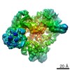
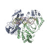
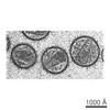
| PubMed Abstract | Reverse transcription of the HIV-1 RNA genome into double-stranded DNA is a central step in viral infection and a common target of antiretroviral drugs . The reaction is catalysed by viral reverse ...Reverse transcription of the HIV-1 RNA genome into double-stranded DNA is a central step in viral infection and a common target of antiretroviral drugs . The reaction is catalysed by viral reverse transcriptase (RT) that is packaged in an infectious virion with two copies of viral genomic RNA each bound to host lysine 3 transfer RNA (tRNA), which acts as a primer for initiation of reverse transcription. Upon viral entry into cells, initiation is slow and non-processive compared to elongation. Despite extensive efforts, the structural basis of RT function during initiation has remained a mystery. Here we use cryo-electron microscopy to determine a three-dimensional structure of an HIV-1 RT initiation complex. In our structure, RT is in an inactive polymerase conformation with open fingers and thumb and with the nucleic acid primer-template complex shifted away from the active site. The primer binding site (PBS) helix formed between tRNA and HIV-1 RNA lies in the cleft of RT and is extended by additional pairing interactions. The 5' end of the tRNA refolds and stacks on the PBS to create a long helical structure, while the remaining viral RNA forms two helical stems positioned above the RT active site, with a linker that connects these helices to the RNase H region of the PBS. Our results illustrate how RNA structure in the initiation complex alters RT conformation to decrease activity, highlighting a potential target for drug action. |
 External links External links | Nature / PubMed:29695867 / PubMed Central |
| Methods | EM (single particle) |
| Resolution | 4.5 - 8.2 Å |
| Structure data | EMDB-7031, PDB-6b19: 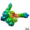 EMDB-7032: 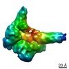 EMDB-7540: |
| Source |
|
 Keywords Keywords |  VIRAL PROTEIN/RNA / Reverse Transcriptase / tRNA / HIV-1 / Reverse Transcription / RNA / Transcription / Complex / RNA-binding protein / backbone model / VIRAL PROTEIN-RNA complex VIRAL PROTEIN/RNA / Reverse Transcriptase / tRNA / HIV-1 / Reverse Transcription / RNA / Transcription / Complex / RNA-binding protein / backbone model / VIRAL PROTEIN-RNA complex |