Movie
Movie Controller
 Structure viewers
 About Yorodumi Papers


+Search query
-Structure paper
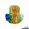
| Title | Domain organization and conformational plasticity of the G protein effector, PDE6. |
|---|---|
| Journal, issue, pages | J Biol Chem, Vol. 290, Issue 20, Page 12833-12843, Year 2015 |
| Publish date | May 15, 2015 |
 Authors Authors | Zhixian Zhang / Feng He / Ryan Constantine / Matthew L Baker / Wolfgang Baehr / Michael F Schmid / Theodore G Wensel / Melina A Agosto /  |
| PubMed Abstract | The cGMP phosphodiesterase of rod photoreceptor cells, PDE6, is the key effector enzyme in phototransduction. Two large catalytic subunits, PDE6α and -β, each contain one catalytic domain and two ...The cGMP phosphodiesterase of rod photoreceptor cells, PDE6, is the key effector enzyme in phototransduction. Two large catalytic subunits, PDE6α and -β, each contain one catalytic domain and two non-catalytic GAF domains, whereas two small inhibitory PDE6γ subunits allow tight regulation by the G protein transducin. The structure of holo-PDE6 in complex with the ROS-1 antibody Fab fragment was determined by cryo-electron microscopy. The ∼11 Å map revealed previously unseen features of PDE6, and each domain was readily fit with high resolution structures. A structure of PDE6 in complex with prenyl-binding protein (PrBP/δ) indicated the location of the PDE6 C-terminal prenylations. Reconstructions of complexes with Fab fragments bound to N or C termini of PDE6γ revealed that PDE6γ stretches from the catalytic domain at one end of the holoenzyme to the GAF-A domain at the other. Removal of PDE6γ caused dramatic structural rearrangements, which were reversed upon its restoration. |
 External links External links | J Biol Chem / PubMed:25809480 / PubMed Central |
| Methods | EM (single particle) |
| Resolution | 11.0 Å |
| Structure data | |
| Chemicals |  ChemComp-IBM: |
| Source |
|
 Keywords Keywords | HYDROLASE/IMMUNE SYSTEM /  phosphodiesterase / photoreceptor / HYDROLASE-IMMUNE SYSTEM complex / PDE6 phosphodiesterase / photoreceptor / HYDROLASE-IMMUNE SYSTEM complex / PDE6 |