Movie
Movie Controller
 Structure viewers
 About Yorodumi Papers


+Search query
-Structure paper
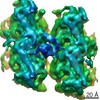
| Title | A structural model for microtubule minus-end recognition and protection by CAMSAP proteins. |
|---|---|
| Journal, issue, pages | Nat Struct Mol Biol, Vol. 24, Issue 11, Page 931-943, Year 2017 |
| Publish date | Oct 9, 2017 |
 Authors Authors | Joseph Atherton / Kai Jiang / Marcel M Stangier / Yanzhang Luo / Shasha Hua / Klaartje Houben / Jolien J E van Hooff / Agnel-Praveen Joseph / Guido Scarabelli / Barry J Grant / Anthony J Roberts / Maya Topf / Michel O Steinmetz / Marc Baldus / Carolyn A Moores / Anna Akhmanova /     |
| PubMed Abstract | CAMSAP and Patronin family members regulate microtubule minus-end stability and localization and thus organize noncentrosomal microtubule networks, which are essential for cell division, polarization ...CAMSAP and Patronin family members regulate microtubule minus-end stability and localization and thus organize noncentrosomal microtubule networks, which are essential for cell division, polarization and differentiation. Here, we found that the CAMSAP C-terminal CKK domain is widely present among eukaryotes and autonomously recognizes microtubule minus ends. Through a combination of structural approaches, we uncovered how mammalian CKK binds between two tubulin dimers at the interprotofilament interface on the outer microtubule surface. In vitro reconstitution assays combined with high-resolution fluorescence microscopy and cryo-electron tomography suggested that CKK preferentially associates with the transition zone between curved protofilaments and the regular microtubule lattice. We propose that minus-end-specific features of the interprotofilament interface at this site serve as the basis for CKK's minus-end preference. The steric clash between microtubule-bound CKK and kinesin motors explains how CKK protects microtubule minus ends against kinesin-13-induced depolymerization and thus controls the stability of free microtubule minus ends. |
 External links External links | Nat Struct Mol Biol / PubMed:28991265 / PubMed Central |
| Methods | EM (single particle) / X-ray diffraction |
| Resolution | 1.4 - 8.0 Å |
| Structure data | EMDB-3444, PDB-5m5c: EMDB-4154, PDB-5m50: EMDB-4156, PDB-5m54:  PDB-5lzn: |
| Chemicals |  ChemComp-HOH: 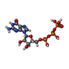 ChemComp-GTP:  ChemComp-MG: 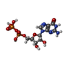 ChemComp-GDP:  ChemComp-TA1: |
| Source |
|
 Keywords Keywords |  STRUCTURAL PROTEIN / -TIP / microtubule / CAMSAP CKK Microtubule Tubulin / MOTOR PROTEIN / CAMSAP / CKK / Tubulin / TRANSPORT PROTEIN STRUCTURAL PROTEIN / -TIP / microtubule / CAMSAP CKK Microtubule Tubulin / MOTOR PROTEIN / CAMSAP / CKK / Tubulin / TRANSPORT PROTEIN |