Movie
Movie Controller
 Structure viewers
 About Yorodumi Papers


+Search query
-Structure paper
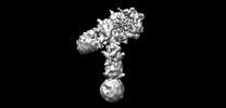
| Title | Molecular basis for the role of disulfide-linked αCTs in the activation of insulin-like growth factor 1 receptor and insulin receptor. |
|---|---|
| Journal, issue, pages | Elife, Vol. 11, Year 2022 |
| Publish date | Nov 22, 2022 |
 Authors Authors | Jie Li / Jiayi Wu / Catherine Hall / Xiao-Chen Bai / Eunhee Choi /  |
| PubMed Abstract | The insulin receptor (IR) and insulin-like growth factor 1 receptor (IGF1R) control metabolic homeostasis and cell growth and proliferation. The IR and IGF1R form similar disulfide bonds linked ...The insulin receptor (IR) and insulin-like growth factor 1 receptor (IGF1R) control metabolic homeostasis and cell growth and proliferation. The IR and IGF1R form similar disulfide bonds linked homodimers in the apo-state; however, their ligand binding properties and the structures in the active state differ substantially. It has been proposed that the disulfide-linked C-terminal segment of α-chain (αCTs) of the IR and IGF1R control the cooperativity of ligand binding and regulate the receptor activation. Nevertheless, the molecular basis for the roles of disulfide-linked αCTs in IR and IGF1R activation are still unclear. Here, we report the cryo-EM structures of full-length mouse IGF1R/IGF1 and IR/insulin complexes with modified αCTs that have increased flexibility. Unlike the -shaped asymmetric IGF1R dimer with a single IGF1 bound, the IGF1R with the enhanced flexibility of αCTs can form a -shaped symmetric dimer with two IGF1s bound. Meanwhile, the IR with non-covalently linked αCTs predominantly adopts an asymmetric conformation with four insulins bound, which is distinct from the -shaped symmetric IR. Using cell-based experiments, we further showed that both IGF1R and IR with the modified αCTs cannot activate the downstream signaling potently. Collectively, our studies demonstrate that the certain structural rigidity of disulfide-linked αCTs is critical for optimal IR and IGF1R signaling activation. |
 External links External links | Elife / PubMed:36413010 / PubMed Central |
| Methods | EM (single particle) |
| Resolution | 3.7 - 4.9 Å |
| Structure data | EMDB-28693, PDB-8eyr: EMDB-28723, PDB-8eyx: EMDB-28724, PDB-8eyy: EMDB-28725, PDB-8ez0: |
| Source |
|
 Keywords Keywords |  SIGNALING PROTEIN / IGF1R / IGF1 / Insulin receptor / insulin SIGNALING PROTEIN / IGF1R / IGF1 / Insulin receptor / insulin |