 Movie
Movie Controller
 Structure viewers
 About Yorodumi Papers


+Search query
-Structure paper
| Title | Structure of the mammalian oligosaccharyl-transferase complex in the native ER protein translocon. |
|---|---|
| Journal, issue, pages | Nat Commun, Vol. 5, Page 3072, Year 2014 |
| Publish date | Nov 24, 2015 |
 Authors Authors | Stefan Pfeffer / Johanna Dudek / Marko Gogala / Stefan Schorr / Johannes Linxweiler / Sven Lang / Thomas Becker / Roland Beckmann / Richard Zimmermann / Friedrich Förster /  |
| PubMed Abstract | In mammalian cells, proteins are typically translocated across the endoplasmic reticulum (ER) membrane in a co-translational mode by the ER protein translocon, comprising the protein-conducting ...In mammalian cells, proteins are typically translocated across the endoplasmic reticulum (ER) membrane in a co-translational mode by the ER protein translocon, comprising the protein-conducting channel Sec61 and additional complexes involved in nascent chain processing and translocation. As an integral component of the translocon, the oligosaccharyl-transferase complex (OST) catalyses co-translational N-glycosylation, one of the most common protein modifications in eukaryotic cells. Here we use cryoelectron tomography, cryoelectron microscopy single-particle analysis and small interfering RNA-mediated gene silencing to determine the overall structure, oligomeric state and position of OST in the native ER protein translocon of mammalian cells in unprecedented detail. The observed positioning of OST in close proximity to Sec61 provides a basis for understanding how protein translocation into the ER and glycosylation of nascent proteins are structurally coupled. The overall spatial organization of the native translocon, as determined here, serves as a reliable framework for further hypothesis-driven studies. |
 External links External links | Nat Commun / PubMed:24407213 |
| Methods | EM (subtomogram averaging) / EM (single particle) |
| Resolution | 9.3 - 40.0 Å |
| Structure data |  EMDB-2514:  EMDB-2515:  EMDB-2516:  EMDB-2517:  EMDB-2518:  EMDB-2519: 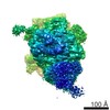 EMDB-2523: |
| Source |
|

 Homo sapiens (human)

