 Movie
Movie Controller
 Structure viewers
 About Yorodumi Papers


+Search query
-Structure paper
| Title | Visualization of two distinct states of disassembly in the bacterial V-ATPase from Thermus thermophilus. |
|---|---|
| Journal, issue, pages | Microscopy (Oxf), Vol. 62, Issue 4, Page 467-474, Year 2013 |
| Publish date | Apr 9, 2013 |
 Authors Authors | Kazutoshi Tani / Christopher P Arthur / Masatada Tamakoshi / Ken Yokoyama / Kaoru Mitsuoka / Yoshinori Fujiyoshi / Christoph Gerle /  |
| PubMed Abstract | V-ATPases are multisubunit, membrane-bound, energy-converting, cellular machines whose assembly and disassembly is innately connected to their activity in vivo. In vitro V-ATPases show a propensity ...V-ATPases are multisubunit, membrane-bound, energy-converting, cellular machines whose assembly and disassembly is innately connected to their activity in vivo. In vitro V-ATPases show a propensity for disassembly that greatly complicates their functional, and, in particular, structural characterization. Direct structural evidence for early stages of their disassembly has not been reported yet. We analyzed the structure of the V-ATPase from Thermus thermophilus in a single negatively stained two-dimensional (2-D) crystal both by electron tomography and by electron crystallography. Our analysis demonstrated that for 2-D crystals of fragile macromolecular complexes, which are too heterogenous or too few for the merging of image data from many crystals, single-crystal 3-D reconstructions by electron tomography and electron crystallography are expedient tools of analysis. The asymmetric unit in the 2-D crystal lattice contains two different V-ATPase complexes that appear to be in an early stage of disassembly and with either one or both peripheral stalks not being visualized, suggesting the involvement of the peripheral stalks in early stages of disassembly. |
 External links External links | Microscopy (Oxf) / PubMed:23572213 |
| Methods | EM (electron crystallography) |
| Resolution | 18.0 Å |
| Structure data | 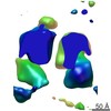 EMDB-2328: |
| Source |
|


 Thermus thermophilus (bacteria)