Movie
Movie Controller
 Structure viewers
 About Yorodumi Papers


+Search query
-Structure paper
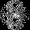
| Title | VASH1-SVBP and VASH2-SVBP generate different detyrosination profiles on microtubules. |
|---|---|
| Journal, issue, pages | J Cell Biol, Vol. 222, Issue 2, Year 2023 |
| Publish date | Feb 6, 2023 |
 Authors Authors | Sacnicte Ramirez-Rios / Sung Ryul Choi / Chadni Sanyal / Thorsten B Blum / Christophe Bosc / Fatma Krichen / Eric Denarier / Jean-Marc Soleilhac / Béatrice Blot / Carsten Janke / Virginie Stoppin-Mellet / Maria M Magiera / Isabelle Arnal / Michel O Steinmetz / Marie-Jo Moutin /   |
| PubMed Abstract | The detyrosination/tyrosination cycle of α-tubulin is critical for proper cell functioning. VASH1-SVBP and VASH2-SVBP are ubiquitous enzymes involved in microtubule detyrosination, whose mode of ...The detyrosination/tyrosination cycle of α-tubulin is critical for proper cell functioning. VASH1-SVBP and VASH2-SVBP are ubiquitous enzymes involved in microtubule detyrosination, whose mode of action is little known. Here, we show in reconstituted systems and cells that VASH1-SVBP and VASH2-SVBP drive the global and local detyrosination of microtubules, respectively. We solved the cryo-electron microscopy structure of VASH2-SVBP bound to microtubules, revealing a different microtubule-binding configuration of its central catalytic region compared to VASH1-SVBP. We show that the divergent mode of detyrosination between the two enzymes is correlated with the microtubule-binding properties of their disordered N- and C-terminal regions. Specifically, the N-terminal region is responsible for a significantly longer residence time of VASH2-SVBP on microtubules compared to VASH1-SVBP. We suggest that this VASH region is critical for microtubule detachment and diffusion of VASH-SVBP enzymes on lattices. Our results suggest a mechanism by which VASH1-SVBP and VASH2-SVBP could generate distinct microtubule subpopulations and confined areas of detyrosinated lattices to drive various microtubule-based cellular functions. |
 External links External links | J Cell Biol / PubMed:36512346 / PubMed Central |
| Methods | EM (single particle) |
| Resolution | 3.6 Å |
| Structure data | EMDB-14634, PDB-7zcw: |
| Chemicals | 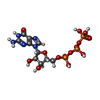 ChemComp-GTP:  ChemComp-MG:  ChemComp-G2P: |
| Source |
|
 Keywords Keywords |  PROTEIN BINDING / Microtubule / Enzyme / Complex / Detyrosination PROTEIN BINDING / Microtubule / Enzyme / Complex / Detyrosination |