 Movie
Movie Controller
 Structure viewers
 About Yorodumi Papers


+Search query
-Structure paper
| Title | Determinants shaping the nanoscale architecture of the mouse rod outer segment. |
|---|---|
| Journal, issue, pages | Elife, Vol. 10, Year 2021 |
| Publish date | Dec 21, 2021 |
 Authors Authors | Matthias Pöge / Julia Mahamid / Sanae S Imanishi / Jürgen M Plitzko / Krzysztof Palczewski / Wolfgang Baumeister /   |
| PubMed Abstract | The unique membrane organization of the rod outer segment (ROS), the specialized sensory cilium of rod photoreceptor cells, provides the foundation for phototransduction, the initial step in vision. ...The unique membrane organization of the rod outer segment (ROS), the specialized sensory cilium of rod photoreceptor cells, provides the foundation for phototransduction, the initial step in vision. ROS architecture is characterized by a stack of identically shaped and tightly packed membrane disks loaded with the visual receptor rhodopsin. A wide range of genetic aberrations have been reported to compromise ROS ultrastructure, impairing photoreceptor viability and function. Yet, the structural basis giving rise to the remarkably precise arrangement of ROS membrane stacks and the molecular mechanisms underlying genetically inherited diseases remain elusive. Here, cryo-electron tomography (cryo-ET) performed on native ROS at molecular resolution provides insights into key structural determinants of ROS membrane architecture. Our data confirm the existence of two previously observed molecular connectors/spacers which likely contribute to the nanometer-scale precise stacking of the ROS disks. We further provide evidence that the extreme radius of curvature at the disk rims is enforced by a continuous supramolecular assembly composed of peripherin-2 (PRPH2) and rod outer segment membrane protein 1 (ROM1) oligomers. We suggest that together these molecular assemblies constitute the structural basis of the highly specialized ROS functional architecture. Our Cryo-ET data provide novel quantitative and structural information on the molecular architecture in ROS and substantiate previous results on proposed mechanisms underlying pathologies of certain PRPH2 mutations leading to blindness. |
 External links External links | Elife / PubMed:34931611 / PubMed Central |
| Methods | EM (subtomogram averaging) |
| Resolution | 19.3 - 29.1 Å |
| Structure data | 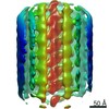 EMDB-13321:  EMDB-13322:  EMDB-13323: 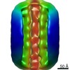 EMDB-13324: |
| Source |
|

 Mus musculus (house mouse)