ムービー
ムービー コントローラー
 構造ビューア
 EMN検索について
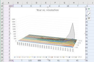


-検索条件
-検索結果
検索 (著者・登録者: jenkins & d)の結果68件中、1から50件目までを表示しています
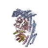
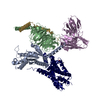
EMDB-19132: 
Structure of dynein-2 intermediate chain DYNC2I2 (WDR34) in complex with dynein-2 heavy chain DYNC2H1.
EMDB-19133:
Structure of dynein-2 intermediate chain DYNC2I1 (WDR60) in complex with the dynein-2 heavy chain DYNC2H1.
PDB-8rgg:
Structure of dynein-2 intermediate chain DYNC2I2 (WDR34) in complex with dynein-2 heavy chain DYNC2H1.
PDB-8rgh:
Structure of dynein-2 intermediate chain DYNC2I1 (WDR60) in complex with the dynein-2 heavy chain DYNC2H1.

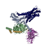
EMDB-41008:
Cryo-EM structure of the DHA bound FFA4-Gq complex

EMDB-41010:
Cryo-EM structure of the Butyrate bound FFA2-Gq complex

EMDB-41013:
Cryo-EM structure of the DHA bound FFA1-Gq complex
PDB-8t3q:
Cryo-EM structure of the DHA bound FFA4-Gq complex

PDB-8t3s:
Cryo-EM structure of the Butyrate bound FFA2-Gq complex

PDB-8t3v:
Cryo-EM structure of the DHA bound FFA1-Gq complex

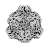
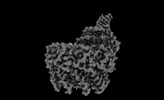
EMDB-43013:
Myxococcus xanthus HEnc-K417N(A) protein shell with icosahedral T=1 symmetry

EMDB-43016:
Myxococcus xanthus HEnc-K417N(A) protein shell with tetrahedral symmetry (12 pentamers, 4 hexamers)
EMDB-43037:
Myxococcus xanthus HEnc-K417N(A) protein shell with D3 symmetry (12 pentamers, 3 hexamers)

EMDB-43038:
Myxococcus xanthus HEnc-K417N(A) protein shell with D6 symmetry (12 pentamers, 8 hexamers)
EMDB-43039:
Myxococcus xanthus HEnc-K417N(A) protein shell with C2 symmetry (12 pentamers, 9 hexamers)
EMDB-43040:
Myxococcus xanthus HEnc-K417N(A) protein shell with D3 symmetry (12 pentamers, 11 hexamers)
EMDB-43041:
Myxococcus xanthus HEnc-K417N(A) protein shell with D2 symmetry (12 pentamers, 12 hexamers)

EMDB-43042:
Myxococcus xanthus HEnc-K417N(A) protein shell with D2 symmetry (12 pentamers, 14 hexamers)
EMDB-43043:
Myxococcus xanthus HEnc-K417N(A) protein shell with D5 symmetry (12 pentamers, 15 hexamers)
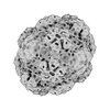
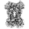
EMDB-43113:
Myxococcus xanthus EncA WT protein shell with icosahedral symmetry T=3

EMDB-41007:
Cryo-EM structure of the TUG-891 bound FFA4-Gq complex
EMDB-41014:
Cryo-EM structure of the DHA bound FFA1-Gq complex(mask on receptor)
PDB-8t3o:
Cryo-EM structure of the TUG-891 bound FFA4-Gq complex

EMDB-43036:
Myxococcus xanthus HEnc-K417N(A) protein shell with icosahedral T=3 symmetry
EMDB-16718:
Drosophila melanogaster insulin receptor ectodomain in complex with DILP5

PDB-8cls:
Drosophila melanogaster insulin receptor ectodomain in complex with DILP5
EMDB-29281:
Cryo-EM structure of STING oligomer bound to cGAMP and NVS-STG2
EMDB-29282:
Cryo-EM structure of STING oligomer bound to cGAMP, NVS-STG2 and C53
PDB-8flk:
Cryo-EM structure of STING oligomer bound to cGAMP and NVS-STG2
PDB-8flm:
Cryo-EM structure of STING oligomer bound to cGAMP, NVS-STG2 and C53
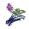
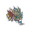
EMDB-29645:
Cryo-EM structure of an orphan GPCR-Gi protein signaling complex
PDB-8g05:
Cryo-EM structure of an orphan GPCR-Gi protein signaling complex
EMDB-17794:
HK97 small terminase in complex with DNA after focused classification

EMDB-17818:
Complex of HK97 small terminase with DNA
PDB-8pop:
HK97 small terminase in complex with DNA

EMDB-27738:
Negative stain EM map of the heterodimeric p110gamma-p84 complex

EMDB-27627:
Structure of p110 gamma bound to the Ras inhibitory nanobody NB7

PDB-8dp0:
Structure of p110 gamma bound to the Ras inhibitory nanobody NB7
EMDB-28658:
Cryo-EM structure of S. aureus BlaR1 with C2 symmetry
EMDB-28659:
Cryo-EM structure of S. aureus BlaR1 with C1 symmetry

EMDB-28660:
Cryo-EM structure of S. aureus BlaR1 TM and zinc metalloprotease domain

EMDB-28661:
Cryo-EM structure of S. aureus BlaR1 F284A mutant

EMDB-28662:
Cryo-EM structure of S. aureus BlaR1 F284A mutant in complex with ampicillin

PDB-8exp:
Cryo-EM structure of S. aureus BlaR1 with C2 symmetry

PDB-8exq:
Cryo-EM structure of S. aureus BlaR1 with C1 symmetry

PDB-8exr:
Cryo-EM structure of S. aureus BlaR1 TM and zinc metalloprotease domain
PDB-8exs:
Cryo-EM structure of S. aureus BlaR1 F284A mutant

PDB-8ext:
Cryo-EM structure of S. aureus BlaR1 F284A mutant in complex with ampicillin

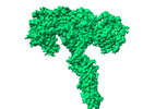
EMDB-27939:
Structure of the human ACE2 receptor in complex with antibody Fab fragment, 05B04

EMDB-25794:
Cryo-EM structure of SARS-CoV-2 spike in complex with antibodies B1-182.1 and A19-61.1
ページ:
 wwPDBはEMDBデータモデルのバージョン3へ移行します